Copyright
©2011 Baishideng Publishing Group Co.
World J Radiol. Apr 28, 2011; 3(4): 114-119
Published online Apr 28, 2011. doi: 10.4329/wjr.v3.i4.114
Published online Apr 28, 2011. doi: 10.4329/wjr.v3.i4.114
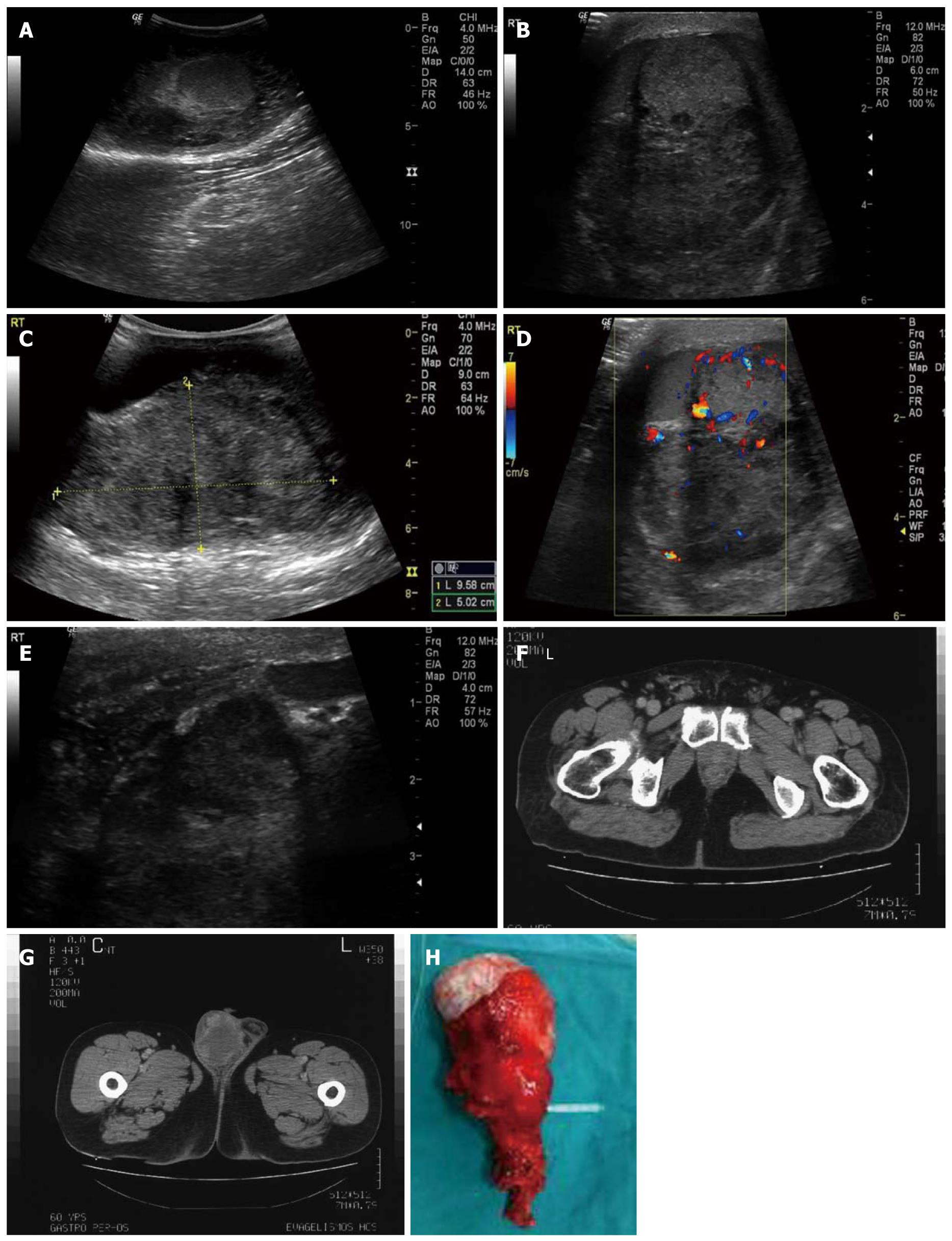
Figure 1 Leiomyosarcoma.
A: Extratesticular mass located posterior-superiorly to the right testis; B: Large mass of heterogenous echotexture located posterior-superiorly to the testis without obvious signs of infiltration; C: Presence of calcifications and hypoechoic areas - hydrocele (use of convex probe due to the size of the mass); D: Colour Doppler image revealing vascularity within the tumour; E: Thickening of the spermatic cord in the inguinal canal; F: Thickening of the spermatic cord and dilated vessels present; G: Mass within the right hemiscrotum with irregular, mostly peripheral vascularity; H: Surgical specimen.
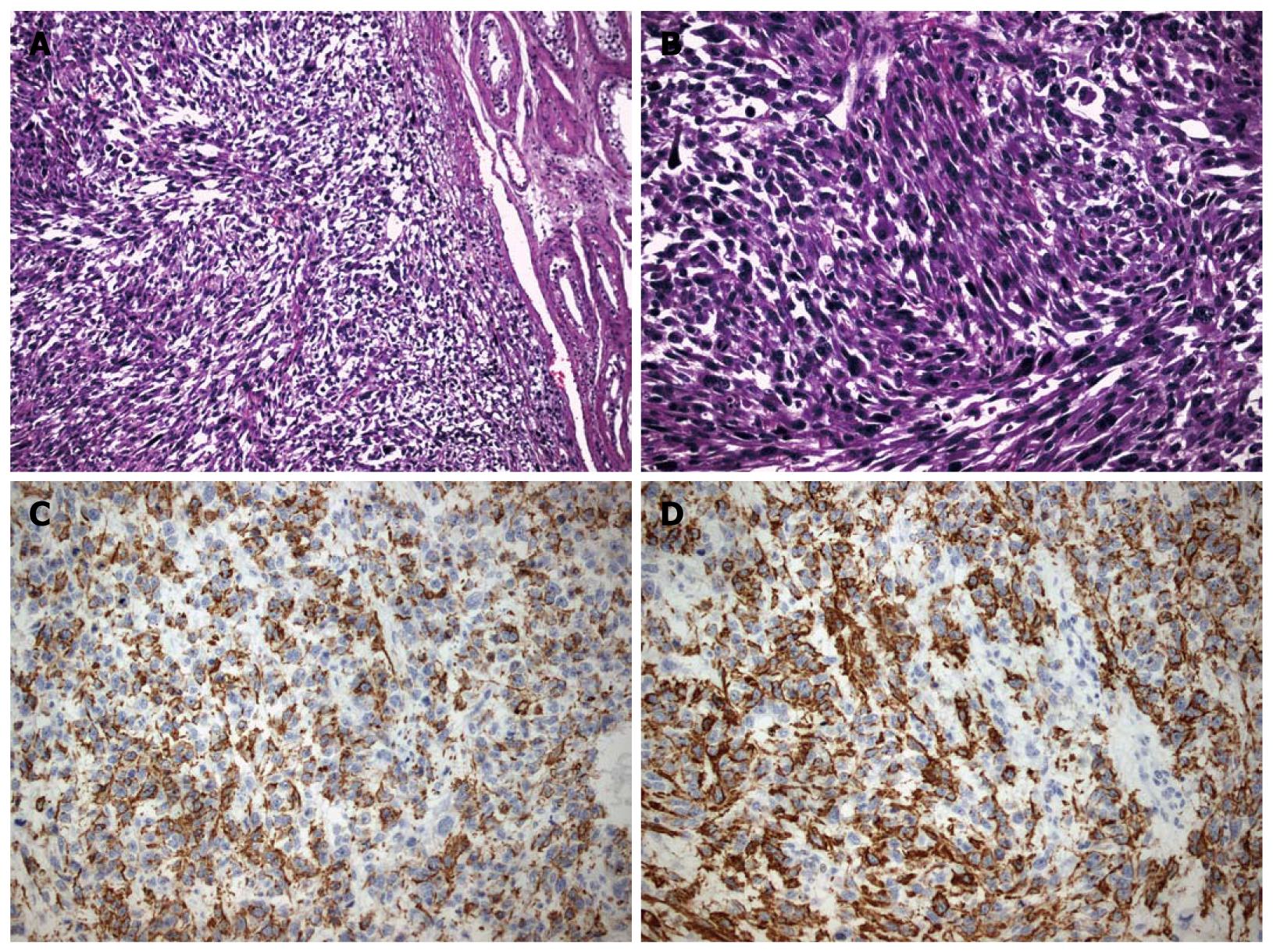
Figure 2 Pathological evidence of spermatic cord leiomyosarcoma.
A: A fascicular growth pattern of spindle neoplastic cells is evident, which is highly indicative of leiomyosarcoma; B: A high degree of atypia and cellular pleomorphism is noticed on the above picture; C: Positivity for desmin is shown; D: Positivity for specific muscle actin is shown.
- Citation: Kyratzi I, Lolis E, Antypa E, Lianou MA, Exarhos D. Imaging features of a huge spermatic cord leiomyosarcoma: Review of the literature. World J Radiol 2011; 3(4): 114-119
- URL: https://www.wjgnet.com/1949-8470/full/v3/i4/114.htm
- DOI: https://dx.doi.org/10.4329/wjr.v3.i4.114


