Copyright
©2011 Baishideng Publishing Group Co.
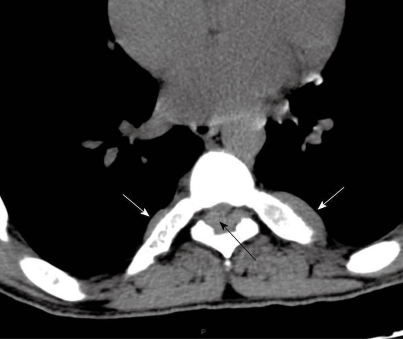
Figure 1 Pre-operative computed tomography scan of the chest without contrast media showing the large bilateral costal masses (white arrows) and an intracanalicular extradural mass (black arrow).
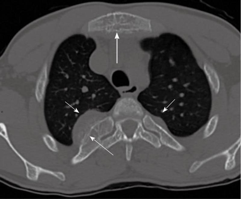
Figure 2 Pre-operative “bone window” computed tomography scan of the chest without contrast media shows large bilateral, well circumscribed lobulated soft tissue masses that cause widening of the ribs (short arrows) and periosteal reaction, without interruption of cortical bone (thin long arrow).
Coexisting involvement of the sternum body (thick long arrow).
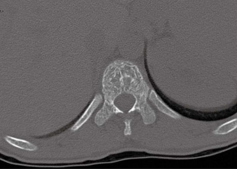
Figure 3 Pre-operative “bone window” computed tomography scan without contrast media: the vertebral body is devoid of bony erosion and has a lacey appearance.
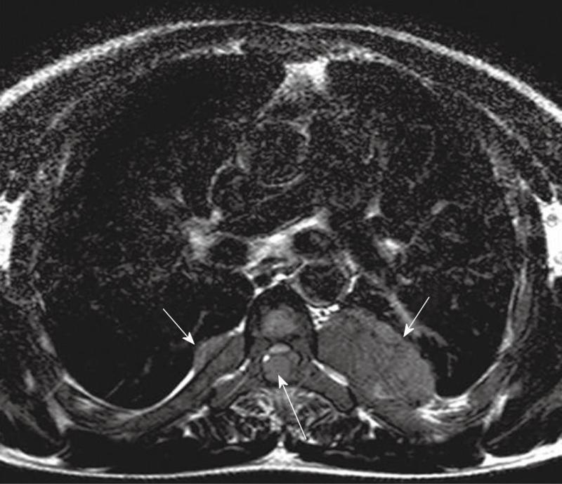
Figure 4 Preoperative axial magnetic resonance imaging, T2 weighted, shows paravertebral and intracanal masses, isointense with the spinal cord, widening the ribs (short arrows) and causing cord compression (long arrow).
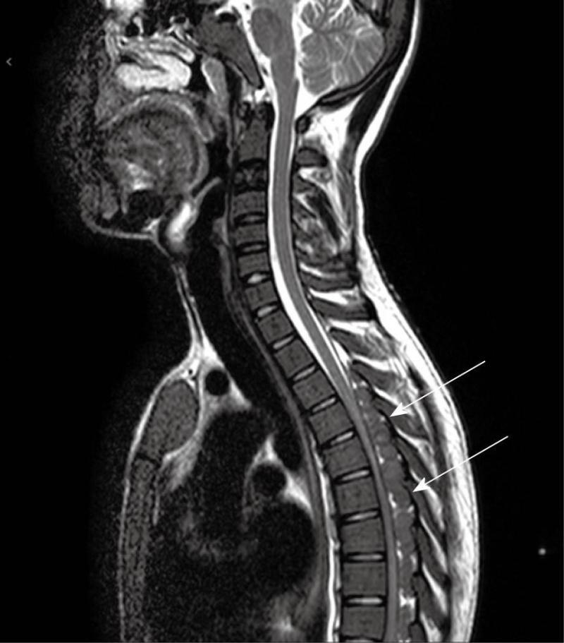
Figure 5 Preoperative sagittal MRI, T2 weighted, shows masses extending from T4 to T9 with cord compression (white arrows), embedded within the epidural fat and isointense with the spinal cord.
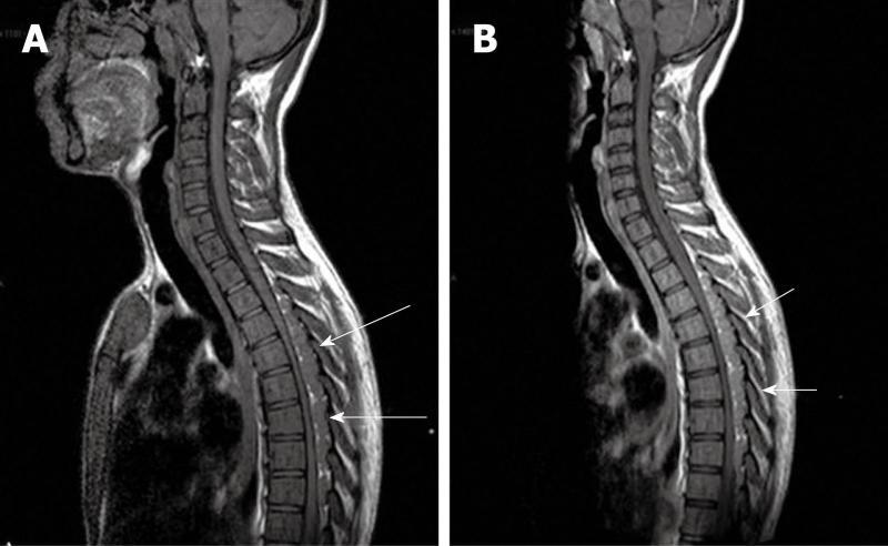
Figure 6 Preoperative sagittal magnetic resonance imaging, T1 weighted, without contrast media (A) (long arrows), and after Gadolinium administration (B) (short arrows) shows poor and homogeneous contrast enhancement of the masses.
- Citation: Savini P, Lanzi A, Marano G, Moretti CC, Poletti G, Musardo G, Foschi FG, Stefanini GF. Paraparesis induced by extramedullary haematopoiesis. World J Radiol 2011; 3(3): 82-84
- URL: https://www.wjgnet.com/1949-8470/full/v3/i3/82.htm
- DOI: https://dx.doi.org/10.4329/wjr.v3.i3.82


