















©2011 Baishideng Publishing Group Co.
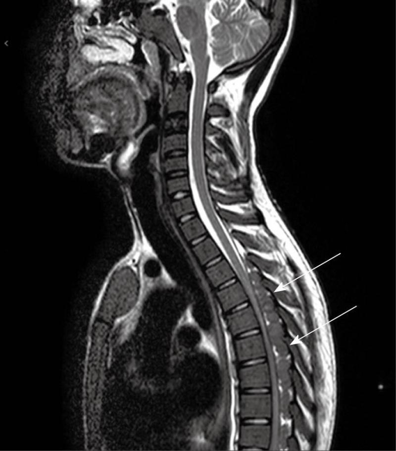
Figure 5 Preoperative sagittal MRI, T2 weighted, shows masses extending from T4 to T9 with cord compression (white arrows), embedded within the epidural fat and isointense with the spinal cord.
- Citation: Savini P, Lanzi A, Marano G, Moretti CC, Poletti G, Musardo G, Foschi FG, Stefanini GF. Paraparesis induced by extramedullary haematopoiesis. World J Radiol 2011; 3(3): 82-84
- URL: https://www.wjgnet.com/1949-8470/full/v3/i3/82.htm
- DOI: https://dx.doi.org/10.4329/wjr.v3.i3.82