Copyright
©2011 Baishideng Publishing Group Co.
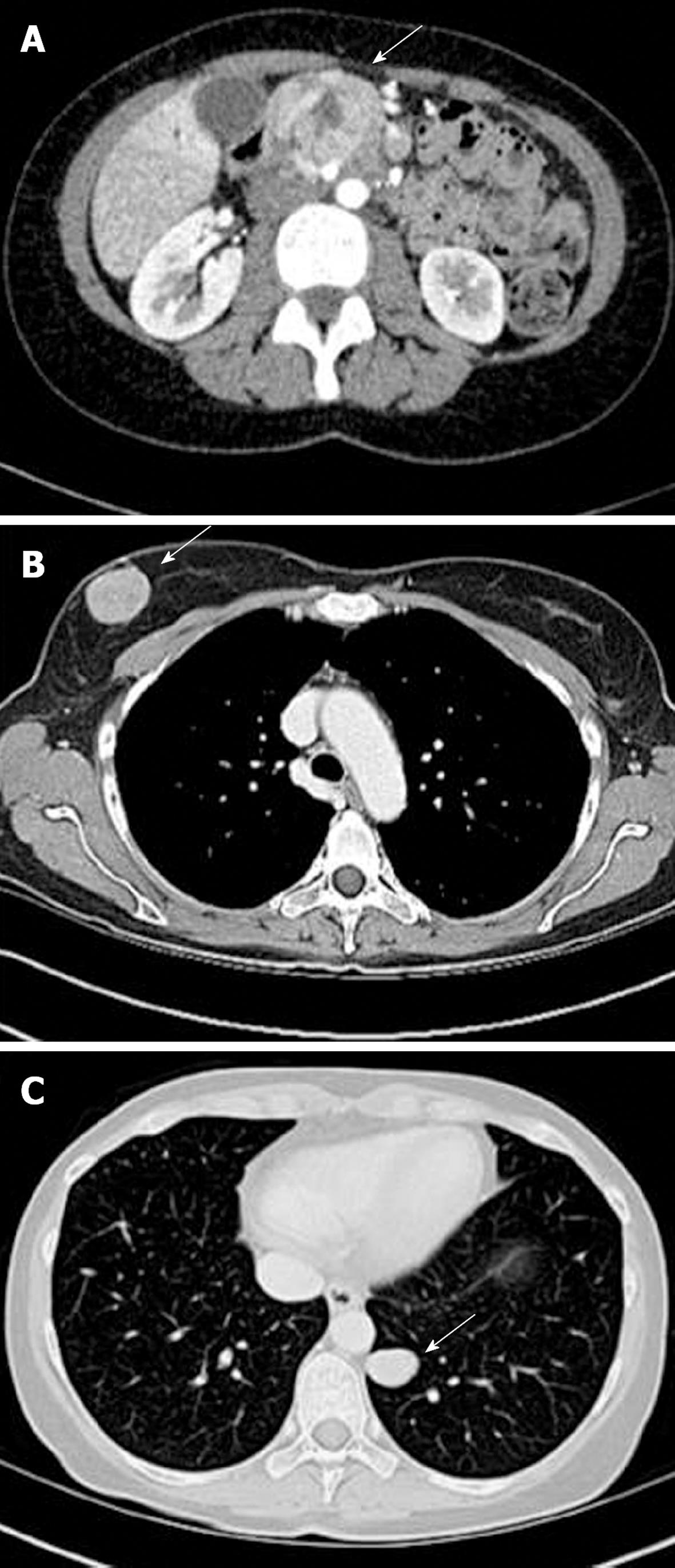
Figure 1 Preoperative computed tomography scans of the chest, abdomen and pelvis.
A: Pancreatic mass with associated biliary dilatation; B: Large, well-circumscribed, right breast lesion; C: Small left lung lesion. Postoperative pathology revealed a poorly differentiated carcinoma with clear cell features, favoring a poorly differentiated neuroendocrine carcinoma in the right breast and left lower lobe of the lung. The arrows depict the lesion of reference.
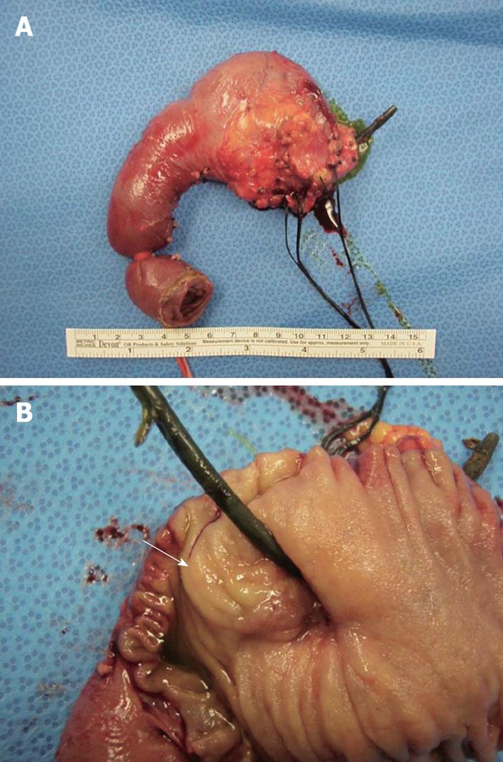
Figure 2 Pyloric sparing Whipple gross specimen with biliary stent in place.
A: There is a marked decrease in the size of the tumor, producing an undetectable remnant lesion on gross examination; B: Small periampullary remnant lesion (arrow) noted on gross examination.
- Citation: Satahoo-Dawes S, Palmer J, III EWM, Levi J. Breast and lung metastasis from pancreatic neuroendocrine carcinoma. World J Radiol 2011; 3(1): 32-37
- URL: https://www.wjgnet.com/1949-8470/full/v3/i1/32.htm
- DOI: https://dx.doi.org/10.4329/wjr.v3.i1.32


