Copyright
©2010 Baishideng Publishing Group Co.
World J Radiol. Sep 28, 2010; 2(9): 374-376
Published online Sep 28, 2010. doi: 10.4329/wjr.v2.i9.374
Published online Sep 28, 2010. doi: 10.4329/wjr.v2.i9.374
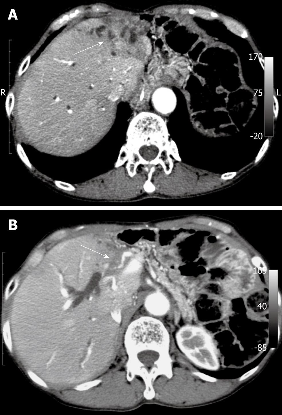
Figure 1 Abdominal computed tomography.
A: Computed tomography computed tomography showing a tumor in the left lobe of the liver (arrow); B: This tumor invading the proper hepatic artery (arrow).
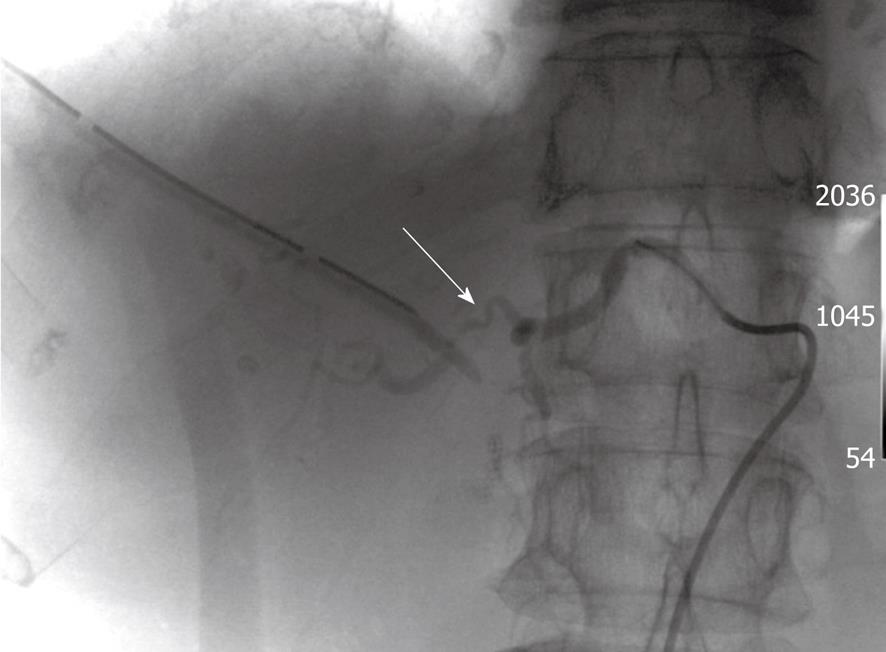
Figure 2 The arterial angiography reveals a fistula between the right hepatic artery and the right hepatic bile duct.
The proximal region of irregular arterial wall of the right hepatic artery is some distance from the biliary catheter (arrow).
- Citation: Hayano K, Miura F, Amano H, Toyota N, Wada K, Kato K, Takada T, Asano T. Arterio-biliary fistula as rare complication of chemoradiation therapy for intrahepatic cholangiocarcinoma. World J Radiol 2010; 2(9): 374-376
- URL: https://www.wjgnet.com/1949-8470/full/v2/i9/374.htm
- DOI: https://dx.doi.org/10.4329/wjr.v2.i9.374


