Copyright
©2010 Baishideng Publishing Group Co.
World J Radiol. Sep 28, 2010; 2(9): 374-376
Published online Sep 28, 2010. doi: 10.4329/wjr.v2.i9.374
Published online Sep 28, 2010. doi: 10.4329/wjr.v2.i9.374
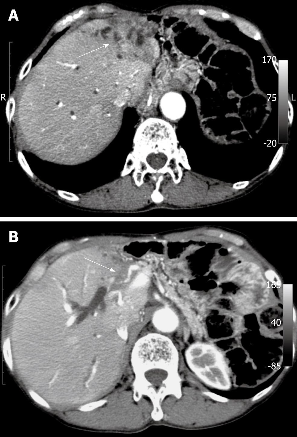
Figure 1 Abdominal computed tomography.
A: Computed tomography computed tomography showing a tumor in the left lobe of the liver (arrow); B: This tumor invading the proper hepatic artery (arrow).
- Citation: Hayano K, Miura F, Amano H, Toyota N, Wada K, Kato K, Takada T, Asano T. Arterio-biliary fistula as rare complication of chemoradiation therapy for intrahepatic cholangiocarcinoma. World J Radiol 2010; 2(9): 374-376
- URL: https://www.wjgnet.com/1949-8470/full/v2/i9/374.htm
- DOI: https://dx.doi.org/10.4329/wjr.v2.i9.374


