Copyright
©2010 Baishideng Publishing Group Co.
World J Radiol. May 28, 2010; 2(5): 151-158
Published online May 28, 2010. doi: 10.4329/wjr.v2.i5.151
Published online May 28, 2010. doi: 10.4329/wjr.v2.i5.151
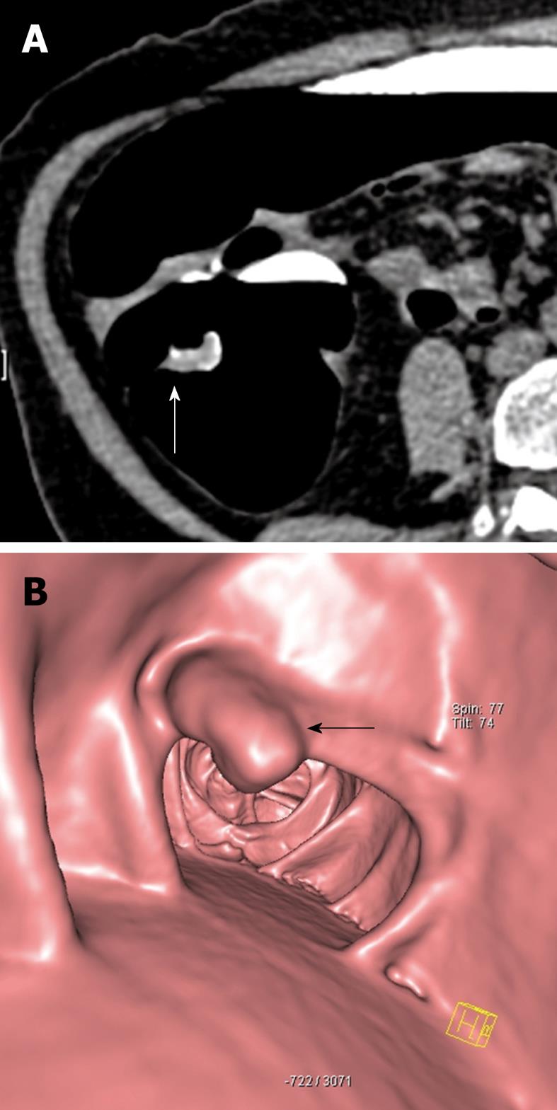
Figure 1 73-year-old female.
Faecal occult blood positive. Sigmoidoscopy was normal. A: Source axial computed tomography (CT) image from CT colonography study in the prone position demonstrates focal thickening (arrow) along a haustral fold in the proximal colon. Note the presence of contrast tagged faecal material coating the lesion; B: 3D reconstructed image of the same lesion showing a polypoid mass (arrow) arising from the haustral fold. Biopsy was positive for adenocarcinoma and the patient underwent curative right hemicolectomy.
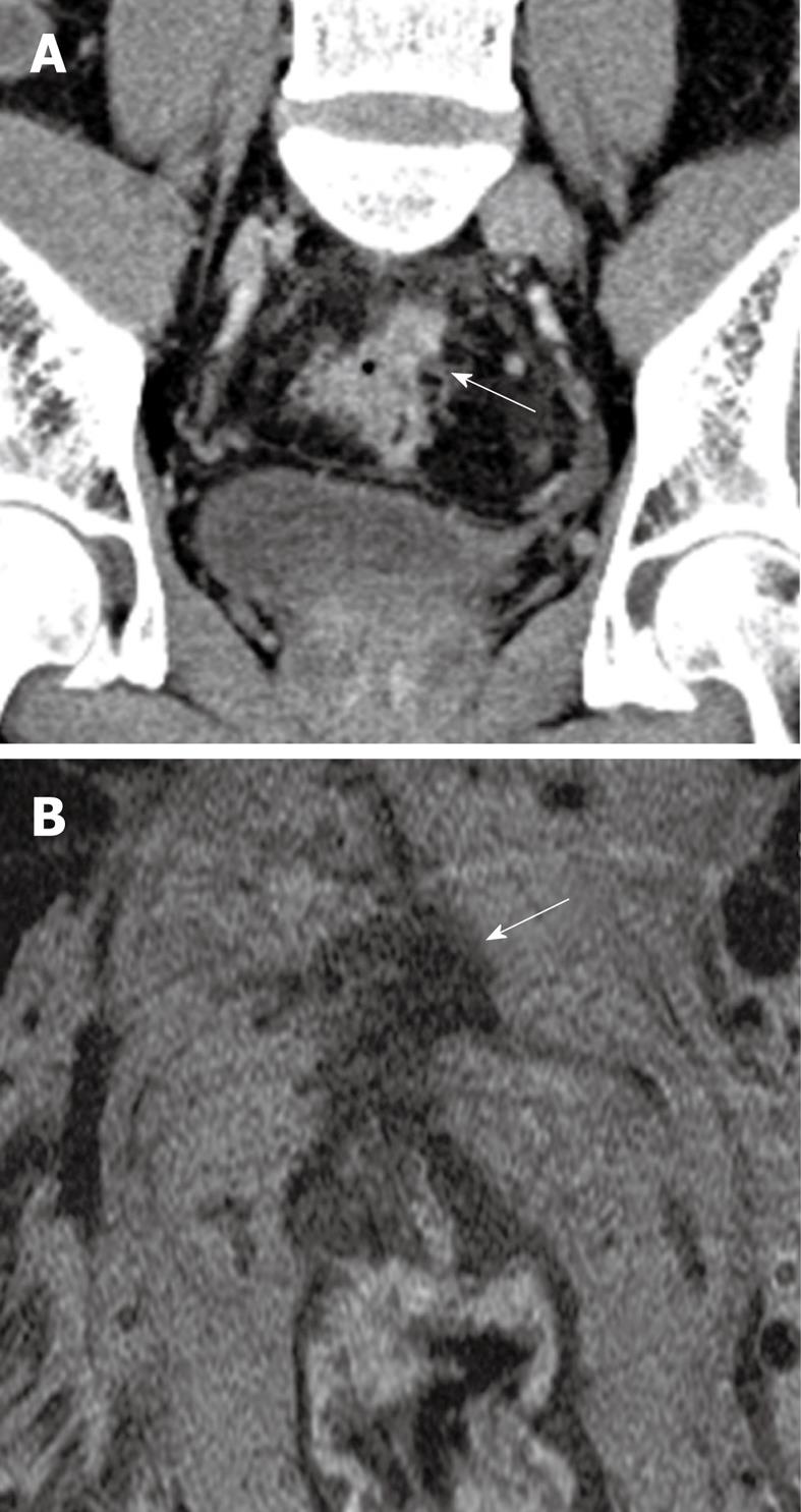
Figure 2 53-year-old male patient.
Presented with lower gastrointestinal bleeding. Endoscopy revealed a fungating mass in the proximal rectum, preventing passage of the scope more proximally. A: Coronal reconstructed contrast-enhanced CT image reveals a spiculated mass at the rectosigmoid junction with a spiculated extraserosal nodular component (arrow); B: Corresponding T2 weighted high resolution magnetic resonance image in the axial plane confirms the findings of extraserosal extension of disease (arrow). The patient was referred for assessment of suitability for neoadjuvant chemoradiation treatment.
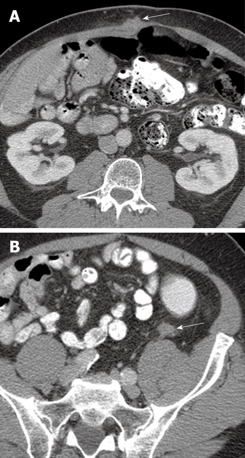
Figure 3 64-year-old male with metastatic adenocarcinoma of the colon.
A: Surveillance axial contrast-enhanced CT image shows a metastatic deposit in the right rectus abdominis muscle (arrow); B: A second metastatic lesion is present in the left paracolic gutter (arrow). The high spatial resolution of CT and the contrast with the adjacent fat allows for easy detection of metastatic disease in these areas.
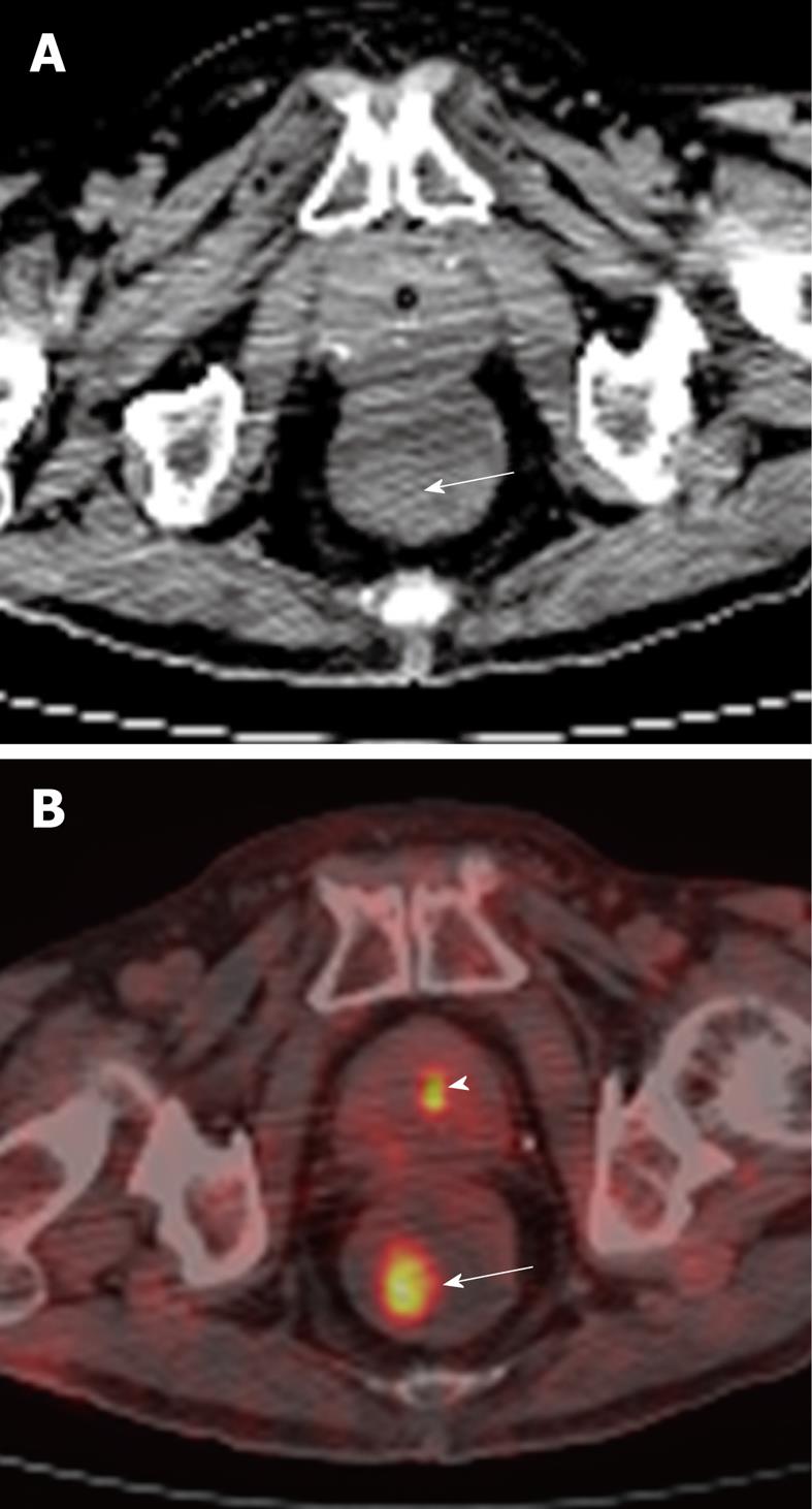
Figure 4 81-year-old male undergoing positron emission tomography (PET)/CT for restaging of diffuse large B cell lymphoma involving the duodenum.
A: Axial non-contrast CT of the rectum showing an incidental subtle soft tissue density polypoid mass (arrow) in the right posterior lateral wall; B: This corresponded to a focal area of hypermetabolic activity (arrow), as demonstrated on fusion PET/CT. Biopsy returned as tubulovillous adenoma. This was excised. Smaller focus of increased tracer activity in keeping with normal physiologic excretion in the urine within the prostatic urethra (arrowhead).
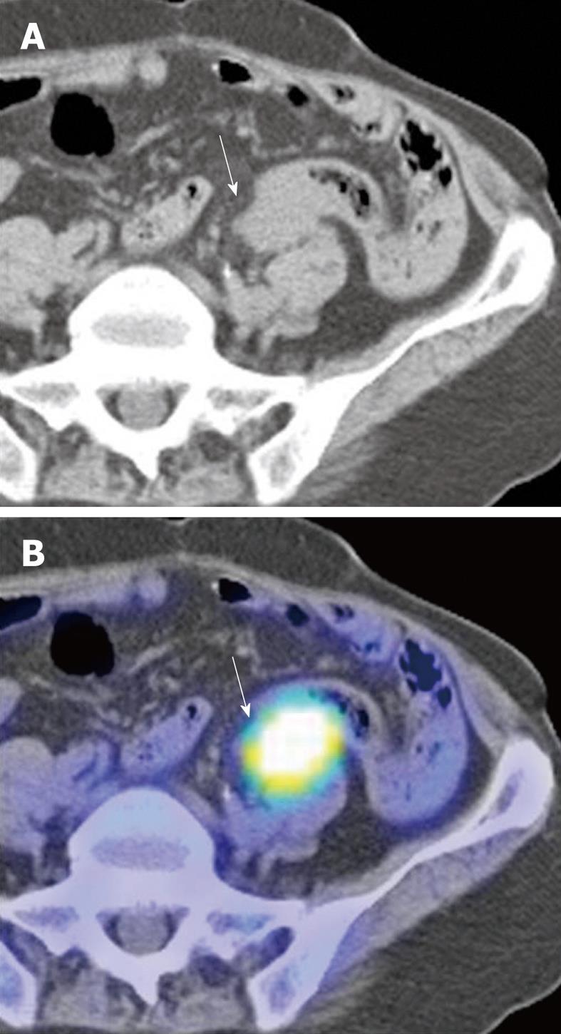
Figure 5 66-year-old Chinese male with adenocarcinoma of the rectum (not shown).
A: Post-operative axial non-contrast CT component of a surveillance PET/CT study showing a soft tissue mass (arrow) abutting the sigmoid colon; B: Corresponding fusion PET/CT image of the lesion (arrow) demonstrates intense hypermetabolic activity consistent with tumour recurrence. (Case courtesy of Dr. Eik Hock Tan, Singapore).
- Citation: Tan CH, Iyer R. Use of computed tomography in the management of colorectal cancer. World J Radiol 2010; 2(5): 151-158
- URL: https://www.wjgnet.com/1949-8470/full/v2/i5/151.htm
- DOI: https://dx.doi.org/10.4329/wjr.v2.i5.151


