Copyright
©2010 Baishideng Publishing Group Co.
World J Radiol. May 28, 2010; 2(5): 151-158
Published online May 28, 2010. doi: 10.4329/wjr.v2.i5.151
Published online May 28, 2010. doi: 10.4329/wjr.v2.i5.151
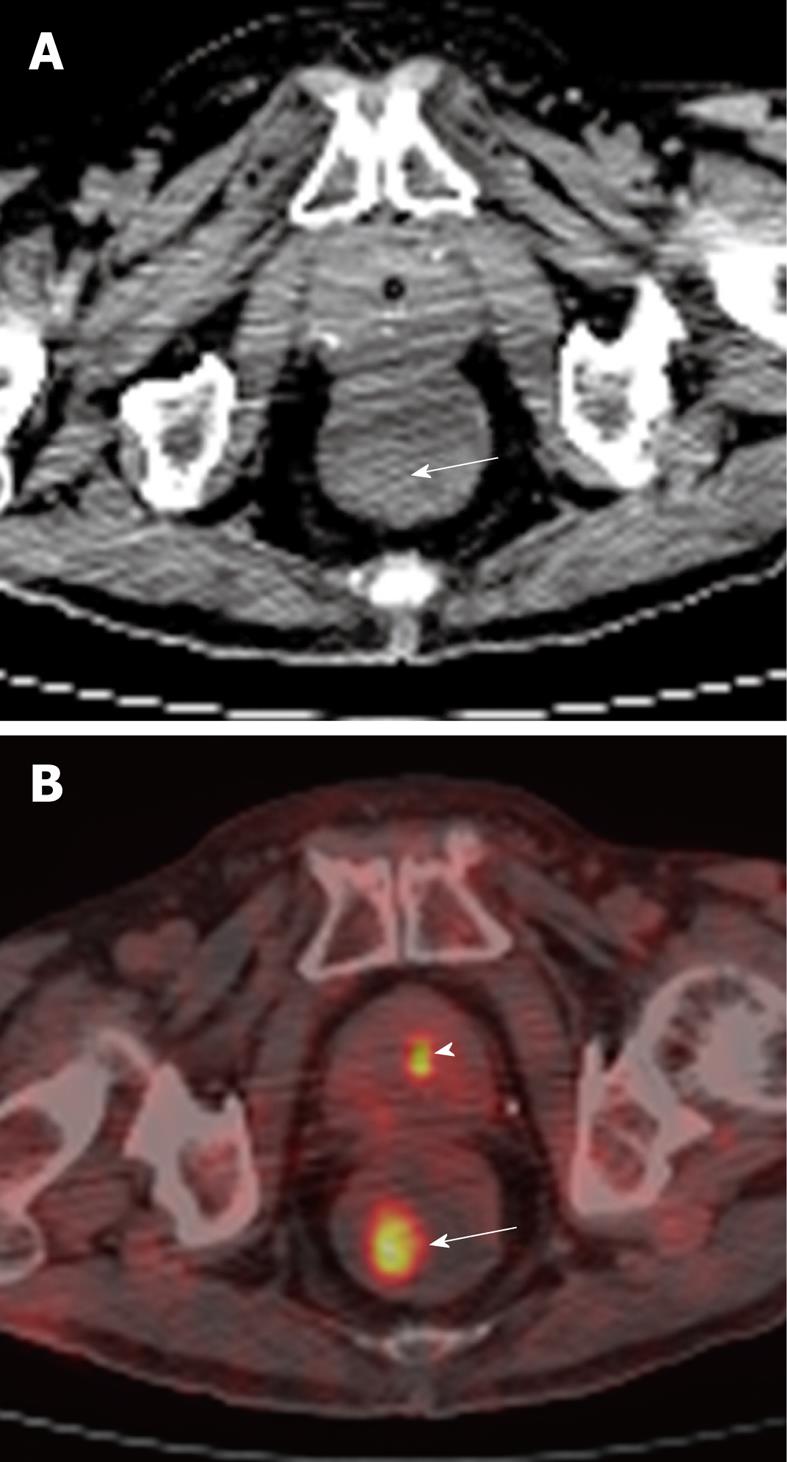
Figure 4 81-year-old male undergoing positron emission tomography (PET)/CT for restaging of diffuse large B cell lymphoma involving the duodenum.
A: Axial non-contrast CT of the rectum showing an incidental subtle soft tissue density polypoid mass (arrow) in the right posterior lateral wall; B: This corresponded to a focal area of hypermetabolic activity (arrow), as demonstrated on fusion PET/CT. Biopsy returned as tubulovillous adenoma. This was excised. Smaller focus of increased tracer activity in keeping with normal physiologic excretion in the urine within the prostatic urethra (arrowhead).
- Citation: Tan CH, Iyer R. Use of computed tomography in the management of colorectal cancer. World J Radiol 2010; 2(5): 151-158
- URL: https://www.wjgnet.com/1949-8470/full/v2/i5/151.htm
- DOI: https://dx.doi.org/10.4329/wjr.v2.i5.151


