Copyright
©2010 Baishideng Publishing Group Co.
World J Radiol. Mar 28, 2010; 2(3): 103-108
Published online Mar 28, 2010. doi: 10.4329/wjr.v2.i3.103
Published online Mar 28, 2010. doi: 10.4329/wjr.v2.i3.103
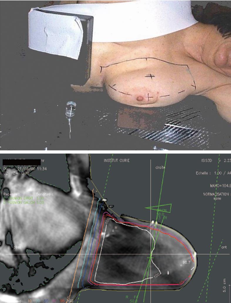
Figure 1 Patient’s position and dosimetry of patient treated in a lateral position.
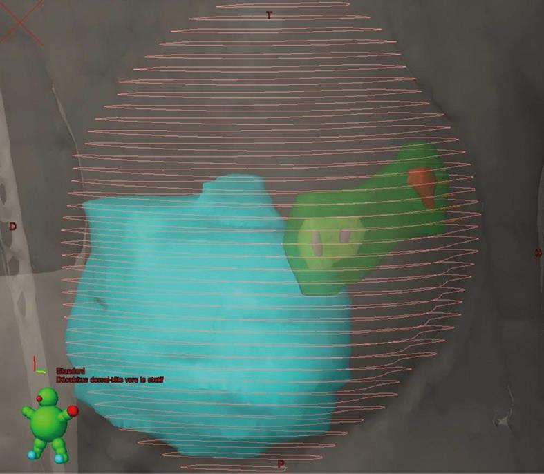
Figure 2 3D reconstruction of boost volume PTV (green) = GTV (red) + CTV clips (yellow), the breast delineation (pink lines) and the relationship between breast volume and boost volume with the cardiac structure[24].
Figure 3 3D reconstruction of defined target volumes and the organs of risk.
A: The left breast is shown in red, heart in pink, lungs in brown and yellow, thyroid in blue, spinal cord in white; B: Process of practical delineation of breast, lymph nodes and organs of risk; C: 3D reconstruction of defined target LN volumes (supra clavicular LN: fuschia, infra clavicular LN: ochre, axilla: dark blue, internal mammary chain: blue, Rotter LN, white) and thyroid (dark blue) as organ of risk.
- Citation: Kirova YM. Recent advances in breast cancer radiotherapy: Evolution or revolution, or how to decrease cardiac toxicity? World J Radiol 2010; 2(3): 103-108
- URL: https://www.wjgnet.com/1949-8470/full/v2/i3/103.htm
- DOI: https://dx.doi.org/10.4329/wjr.v2.i3.103


