Copyright
©The Author(s) 2025.
World J Radiol. Jun 28, 2025; 17(6): 107315
Published online Jun 28, 2025. doi: 10.4329/wjr.v17.i6.107315
Published online Jun 28, 2025. doi: 10.4329/wjr.v17.i6.107315
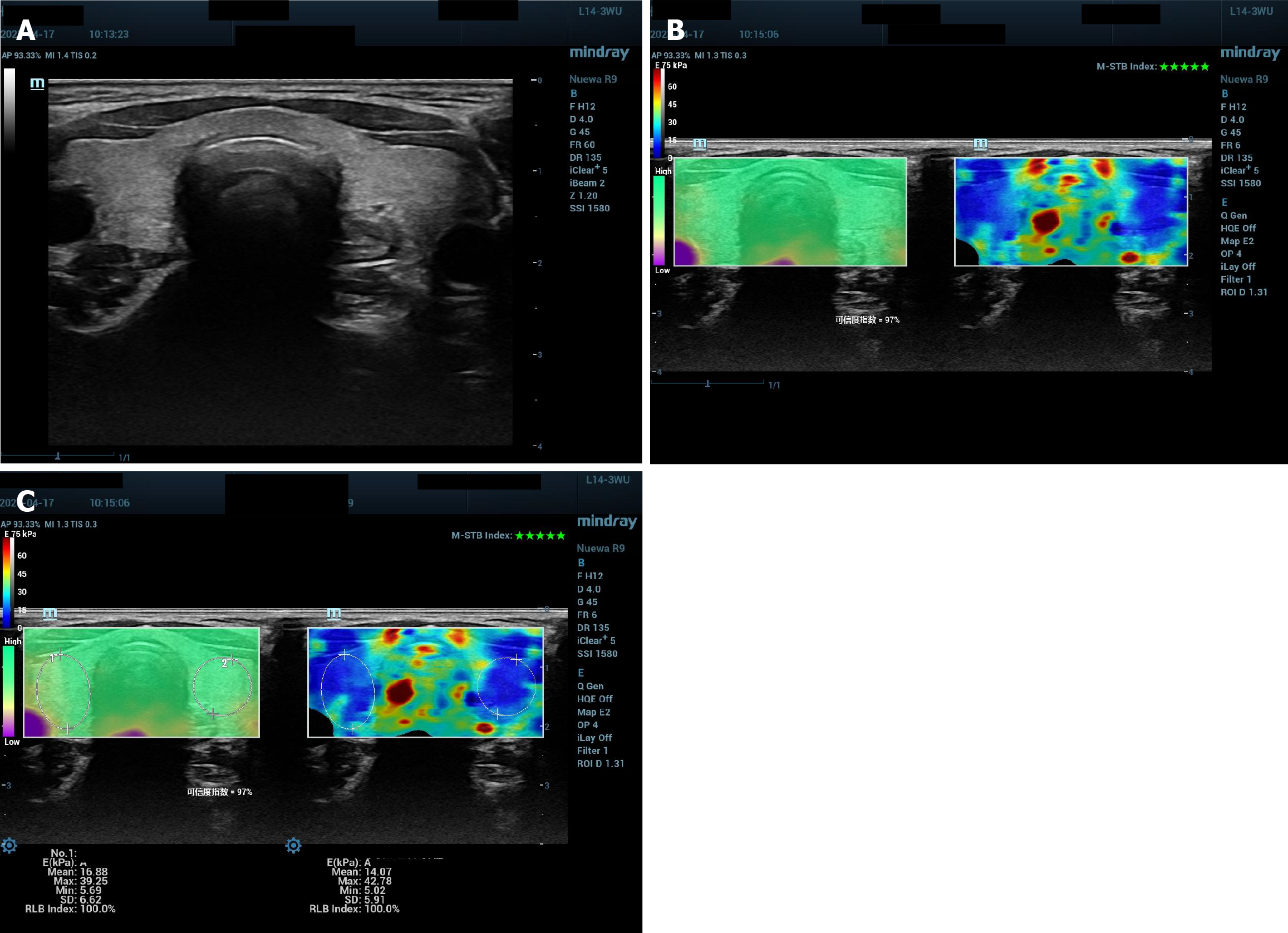
Figure 1 Thyroid ultrasonography and two-dimensional shear wave elastography: normal morphology and elasticity assessment with quality-controlled imaging.
A: Conventional ultrasound scan of transverse section of thyroid, with normal size and homogeneous isoechogenicity; B: Two-dimensional shear wave elastography (2D SWE) image of thyroid transverse section met quality control criteria. The right was 2D SWE image with blue as soft and red as hard; five stars in the top right-hand corner suggested with perfect motion-stability. The left image showed the reliability of 2D SWE image, with green as good reliability, purple as poor and reliability index as 97%; C: Region of interest (ROI) drawing shown on Figure 1B. An oval ROI was drawn to include the right lobe; another oval ROI was drawn to include the left lobe. Quantitative parameters as mean elasticity in the ROI and maximal elasticity in the ROI shown on the screen were recorded for further analysis.
- Citation: Zhang HP, Chen ML, Zou J, Zhou YQ. Value of two-dimensional shear wave elastography quantitative analysis for evaluation of thyroid function in first trimester pregnancy. World J Radiol 2025; 17(6): 107315
- URL: https://www.wjgnet.com/1949-8470/full/v17/i6/107315.htm
- DOI: https://dx.doi.org/10.4329/wjr.v17.i6.107315


