Copyright
©The Author(s) 2021.
World J Radiol. Jan 28, 2021; 13(1): 19-28
Published online Jan 28, 2021. doi: 10.4329/wjr.v13.i1.19
Published online Jan 28, 2021. doi: 10.4329/wjr.v13.i1.19
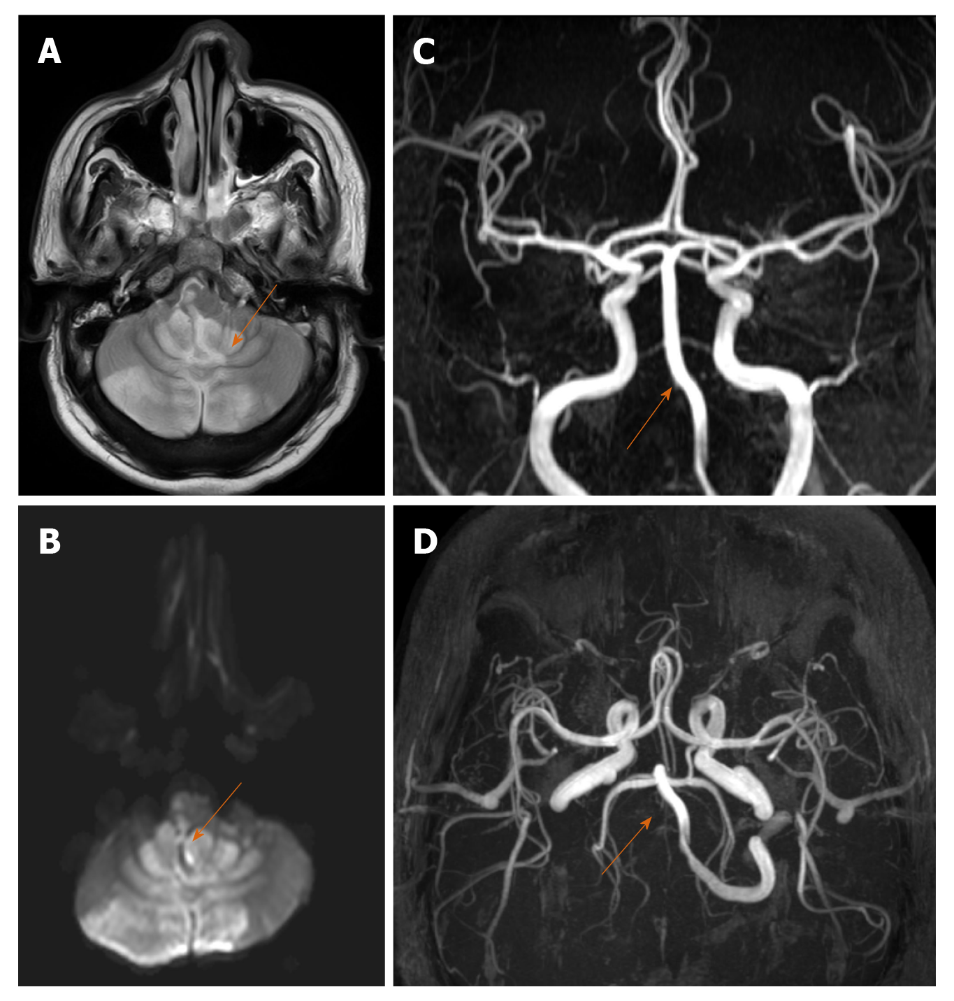
Figure 1 A 35-year-old male with posterior circulation stroke.
A and B: Axial sections of magnetic resonance imaging brain (T2W, diffusion sequences) show areas of high signal in both cerebellar hemispheres, vermis and brainstem suggestive of acute infarcts; C and D: Magnetic resonance artery coronal and axial sections show complete non visualization of right vertebral artery (arrow) suggestive of thrombosis.
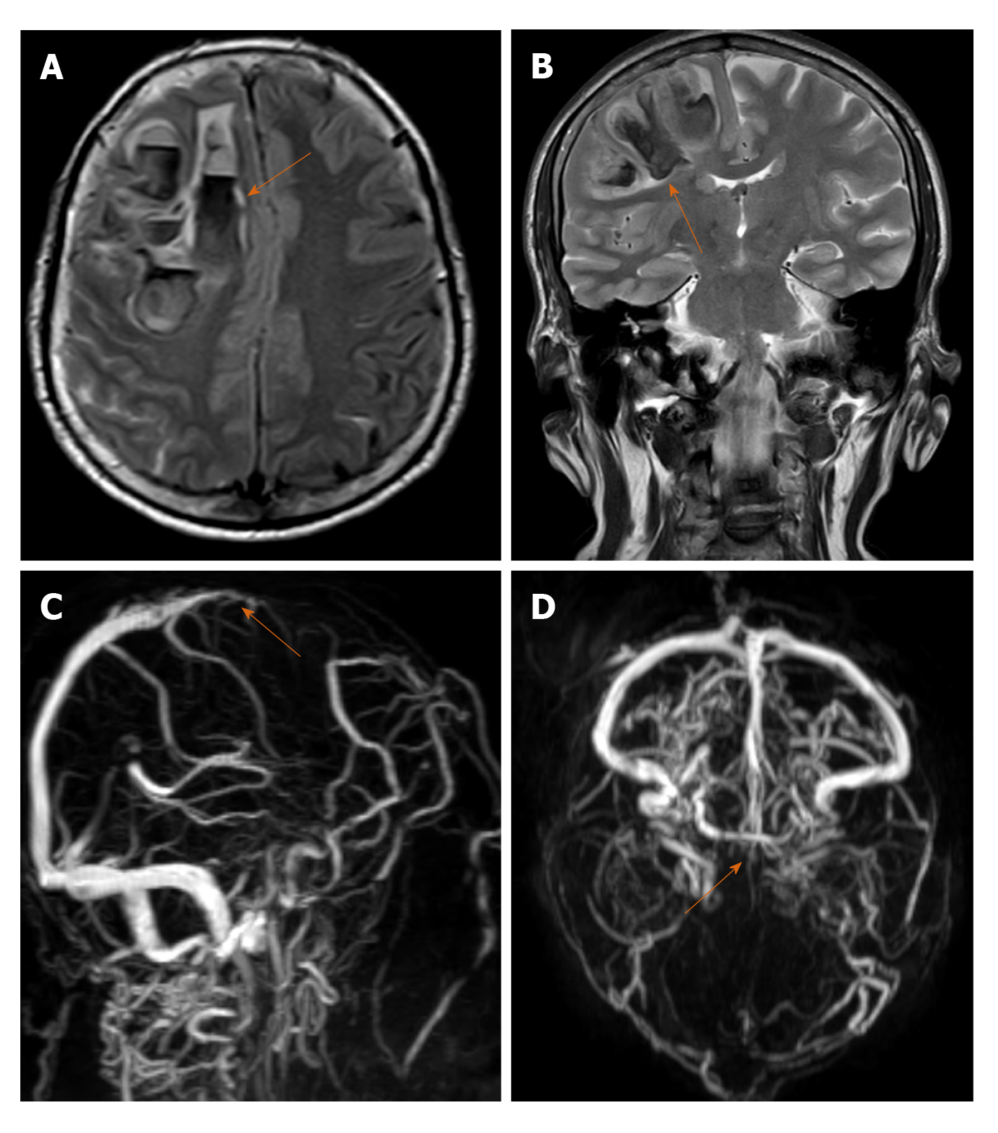
Figure 2 A 61-year-old male with Dural venous thrombosis.
A and B: Axial and coronal sections of magnetic resonance imaging brain (T2WI sequence) show acute hemorrhage (arrow) in right frontal lobe with left sided midline shift; C and D: Magnetic resonance venography sagittal oblique and axial sections show absent flow related signal in anterior third of superior sagittal sinus suggestive of thrombosis.
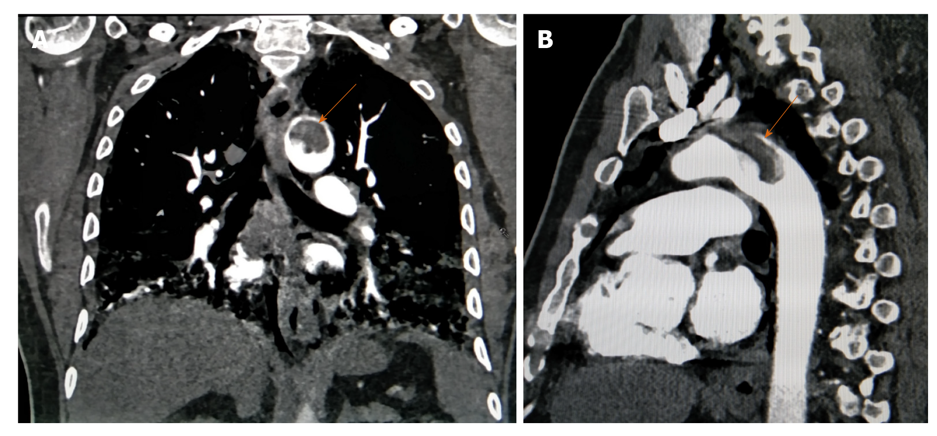
Figure 3 A 64-year-old male with aortic mural thrombosis.
A and B: Coronal and sagittal sections of arterial phase of contrast-enhanced computed tomography thorax show pedunculated thrombus in aortic arch suggestive of aortic mural thrombus.
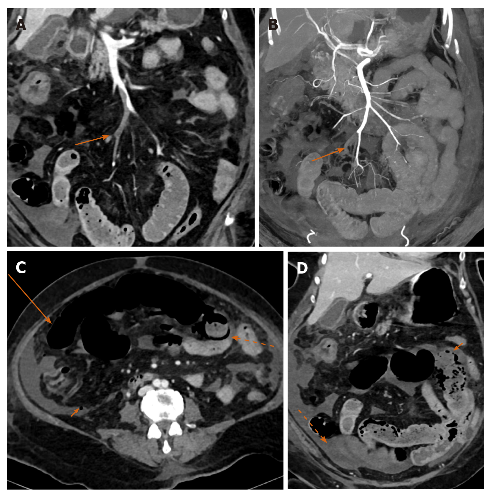
Figure 4 A 65-year-old female with acute mesenteric ischemia.
A: Coronal reformatted image of contrast-enhanced computed tomography (CECT) abdomen shows filling defects (orange arrow) in ileal branches of superior mesenteric artery suggestive of thrombosis; B: Coronal reformatted image of CECT abdomen shows occlusion of accompanying tributaries of superior mesenteric vein (SMV) with superior extension of thrombus into the main stem of SMV; C: Axial CECT image showing dilated small bowel with paper thin wall (long orange arrow), circumferential pneumatosis (dotted orange arrow) and foci of free extraluminal air (small orange arrow) indicating transmural bowel necrosis with perforation; D: Coronal CECT image showing a bowel segment with absent mural enhancement (solid orange arrow), and ascites (dotted arrow).
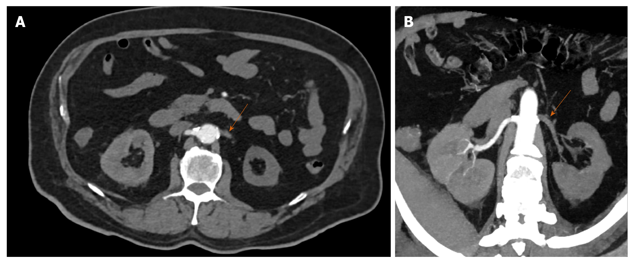
Figure 5 A 74-year-old male with renal artery thrombosis.
A and B: Axial baseline and oblique coronal reformatted maximum intensity projection images of arterial phase contrast-enhanced computed tomography images showing hypodense filling defect involving left renal artery from ostium to hilum and its segmental branches with non-enhancement of left kidney suggestive of left renal artery thrombosis with infarct.
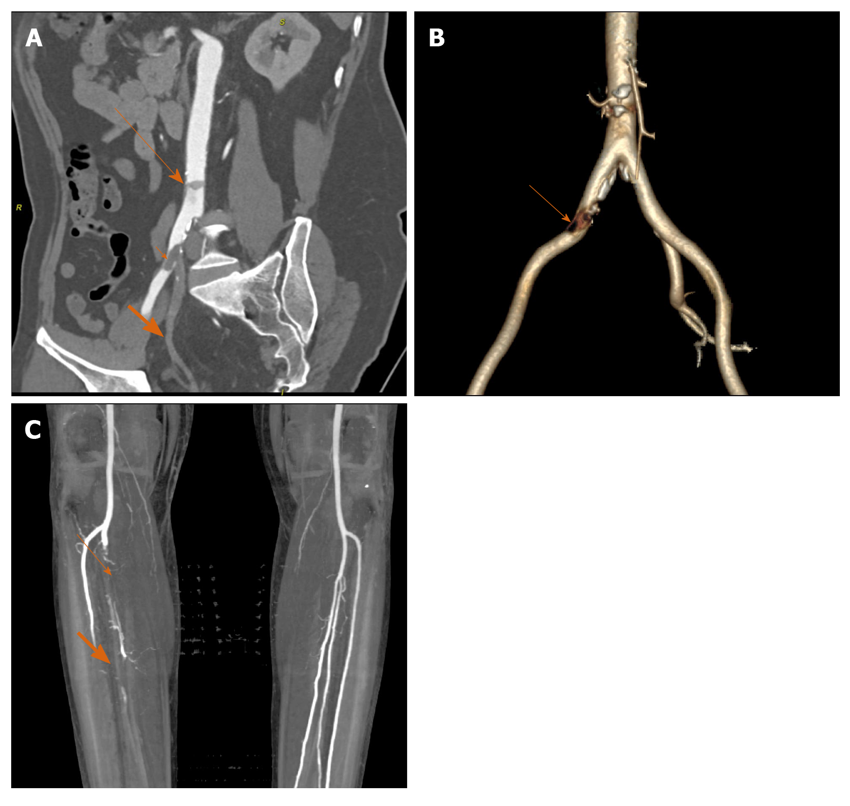
Figure 6 A 51-year-old male with peripheral arterial disease.
A: Coronal oblique reformatted image of contrast-enhanced computed tomography abdomen shows mall mural thrombus in abdominal aorta (long arrow); and another partially occluding thrombus at right common iliac artery (short arrow) bifurcation extending into external iliac branch and synchronous complete thrombosis of right internal iliac artery (broad orange arrow); B: Three-dimensional reconstructed image shows defect in right common iliac artery and complete non visualization of right internal iliac artery; C: Coronal maximum intensity projection image of computed tomography angiography of bilateral lower limbs shows filling defect in right tibio-peroneal trunk just beyond origin with poor distal reformation (thin arrow), and non-opacification of mid and distal third of right anterior tibial artery (broad orange arrow).
- Citation: Shankar A, Varadan B, Ethiraj D, Sudarsanam H, Hakeem AR, Kalyanasundaram S. Systemic arterio-venous thrombosis in COVID-19: A pictorial review. World J Radiol 2021; 13(1): 19-28
- URL: https://www.wjgnet.com/1949-8470/full/v13/i1/19.htm
- DOI: https://dx.doi.org/10.4329/wjr.v13.i1.19


