Copyright
©The Author(s) 2020.
World J Radiol. Dec 28, 2020; 12(12): 289-301
Published online Dec 28, 2020. doi: 10.4329/wjr.v12.i12.289
Published online Dec 28, 2020. doi: 10.4329/wjr.v12.i12.289
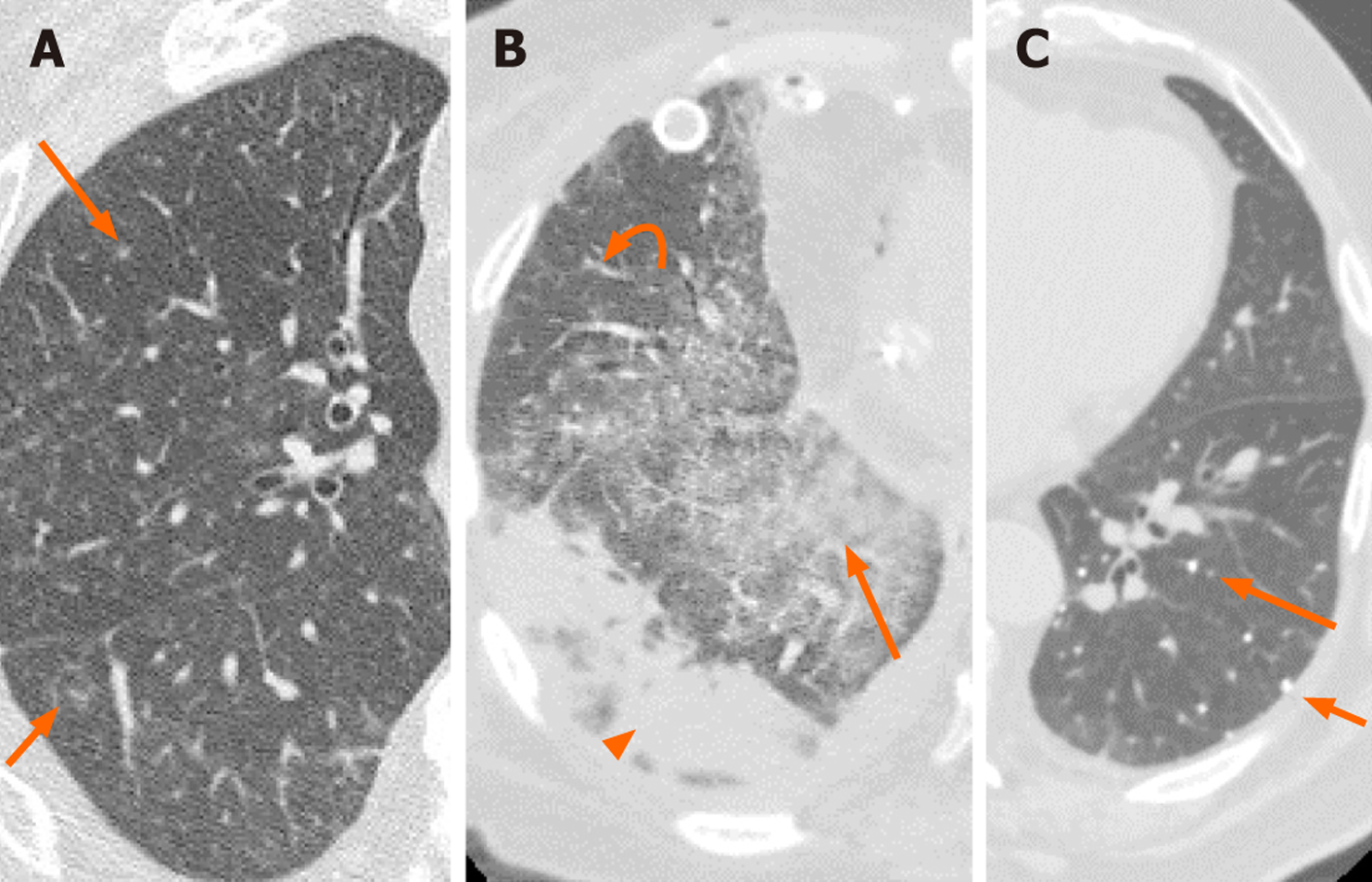
Figure 1 Two patients with double lung transplants and cytomegalovirus pneumonia diagnosed via transbronchial biopsy and lavage respectively.
A: Axial chest computed tomography (CT) in a 67-year-old male shows numerous nodular infiltrates (arrows); B: The CT for a 56-year-old female shows ground glass opacities (arrow), reticular opacities (curved arrow) and sub pleural consolidation (arrowhead); C: The third patient is a 76-year-old male with a history of varicella pneumonia. There are numerous small calcified nodules (arrows). However, this appearance can also be seen in pulmonary hemosiderosis, Goodpasture syndrome, silicosis, pulmonary alveolar microlithiasis, and calcified metastasis.
Figure 2 A 37-year-old female with a history of double lung transplant presented with progressive dyspnea.
Axial computed tomography of the chest demonstrates bronchocentric multifocal opacities (black circles; A-C). Transbronchial lung biopsy demonstrated herpes simplex virus pneumonia.
Figure 3 A 33-year-old man with hypoxic respiratory failure, requiring intubation was polymerase chain reaction positive for influenza A.
A: Radiograph shows diffuse granular opacities resembling pulmonary interstitial edema; B: A contemporaneous axial computed tomography (CT) (arrows) shows diffuse ground glass opacities with patchy consolidations that are both in peripheral and central zones. Pleural effusion (asterisk) is more common in influenza A pneumonia than coronavirus disease 2019 (COVID-19); C: Axial CT two months later demonstrates cavitation (arrow) and traction bronchiectasis (curved arrow) which are not frequently reported in COVID-19.
Figure 4 A 72-year-old man presented with fever, dry cough and shortness of breath.
Polymerase chain reaction was positive for severe acute respiratory syndrome coronavirus 2 infection. A and B: Axial chest computed tomography shows bilateral peripheral ground glass opacities (arrows; A and B) and superimposed reticulation (arrowhead; B), consistent with coronavirus disease 2019 pneumonia.
Figure 5 A 46-year-old woman presented with fever and dry cough.
Polymerase chain reaction was positive for severe acute respiratory syndrome coronavirus 2 infection. A and B: Axial chest computed tomography shows bilateral multifocal ground glass opacities (arrows; A and B), peribronchial interstitial thickening (arrowhead; B) and reticular opacities (curved arrows; B), consistent with coronavirus disease 2019 pneumonia.
Figure 6 A 35-year-old previously healthy woman presented with fever, dry cough and shortness of breath 10 d after contact with a sick person with coronavirus disease 2019.
A: Radiograph shows haziness (arrows) in basilar portions; B: Axial chest computed tomography (CT) in the same day demonstrates bilateral multifocal ground glass opacities (GGOs) (arrows) in favor of coronavirus disease 2019 infection that was confirmed with positive reverse transcriptase polymerase chain reaction. The patient’s condition worsened and she was intubated; C: Follow-up CT after 3 d shows progression of GGOs (arrows), superimposed consolidations (arrowheads), and ill-defined nodular opacities (thin arrows). Ultimately, her condition improved and she was discharged in a good condition after 20 d.
- Citation: Eslambolchi A, Maliglig A, Gupta A, Gholamrezanezhad A. COVID-19 or non-COVID viral pneumonia: How to differentiate based on the radiologic findings? World J Radiol 2020; 12(12): 289-301
- URL: https://www.wjgnet.com/1949-8470/full/v12/i12/289.htm
- DOI: https://dx.doi.org/10.4329/wjr.v12.i12.289


