
















©The Author(s) 2020.
World J Radiol. Dec 28, 2020; 12(12): 289-301
Published online Dec 28, 2020. doi: 10.4329/wjr.v12.i12.289
Published online Dec 28, 2020. doi: 10.4329/wjr.v12.i12.289
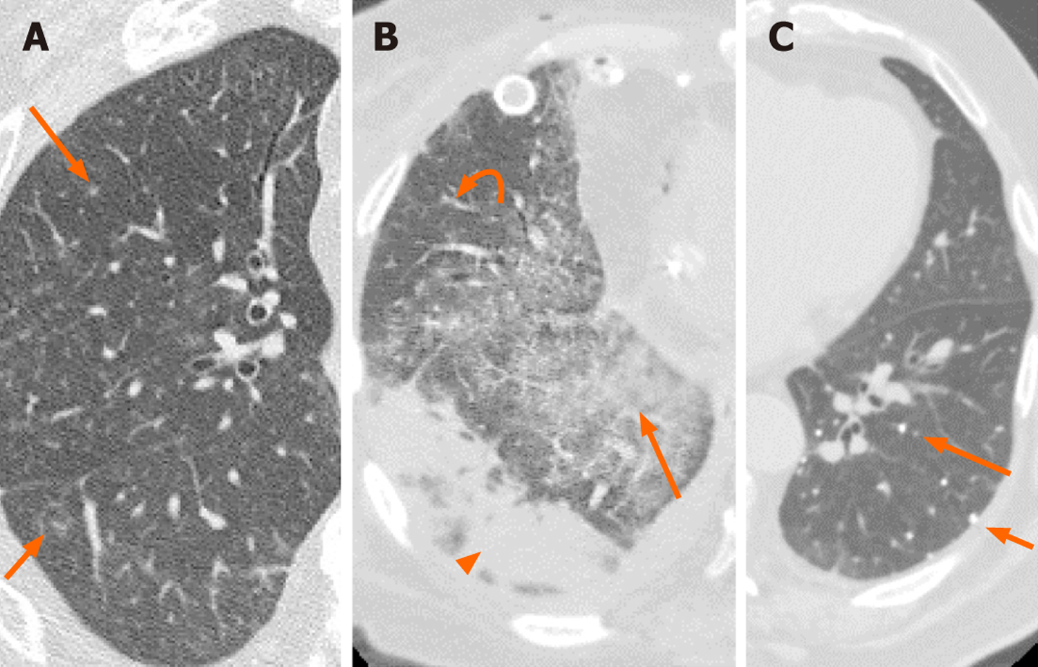
Figure 1 Two patients with double lung transplants and cytomegalovirus pneumonia diagnosed via transbronchial biopsy and lavage respectively.
A: Axial chest computed tomography (CT) in a 67-year-old male shows numerous nodular infiltrates (arrows); B: The CT for a 56-year-old female shows ground glass opacities (arrow), reticular opacities (curved arrow) and sub pleural consolidation (arrowhead); C: The third patient is a 76-year-old male with a history of varicella pneumonia. There are numerous small calcified nodules (arrows). However, this appearance can also be seen in pulmonary hemosiderosis, Goodpasture syndrome, silicosis, pulmonary alveolar microlithiasis, and calcified metastasis.
- Citation: Eslambolchi A, Maliglig A, Gupta A, Gholamrezanezhad A. COVID-19 or non-COVID viral pneumonia: How to differentiate based on the radiologic findings? World J Radiol 2020; 12(12): 289-301
- URL: https://www.wjgnet.com/1949-8470/full/v12/i12/289.htm
- DOI: https://dx.doi.org/10.4329/wjr.v12.i12.289