Copyright
©The Author(s) 2018.
World J Radiol. Oct 28, 2018; 10(10): 116-123
Published online Oct 28, 2018. doi: 10.4329/wjr.v10.i10.116
Published online Oct 28, 2018. doi: 10.4329/wjr.v10.i10.116
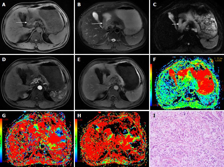
Figure 1 A 48-year-old male diagnosed with malignant gastric carcinoma (signet ring cell cancer).
A, B: The lesion has slightly low signal on T1-weighted image (A) and slightly high signal intensity on T2-weighted image (B); C: On DWI, the cancer shows hyperintensity (white arrows); D, E: After contrast agent injection, the lesion shows mild-to-moderate enhancement in arterial and portal venous phases; F-H: The pseudocolor maps of D, D* and f derived from IVIM were displayed, the values of the D, D* and f are 0.92 ± 0.11 × 10⁻³ mm2/s, 26.75 ± 13.61 × 10⁻³ mm2/s and 17.24% ± 4.8%, respectively; I: The HE staining of the tissues (100 ×).
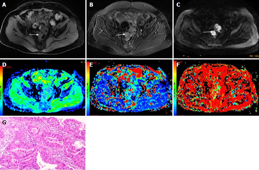
Figure 2 A 67-year-old female diagnosed as rectal cancer (poorly differentiated adenocarcinoma).
A, B: The rectal cancer is isointense on T1-weighted image (A) with slightly high signal intensity on T2-weighted image (B); C: On diffusion weighted imaging, the cancer shows hyperintensity (white arrows); D-F: The pseudocolor maps of D, D* and f derived from intravoxel incoherent motion are displayed, the values of the D, D* and f were 1.03 ± 0.12 × 10⁻³mm2/s, 50.35 ± 24.96 × 10⁻³mm2/s and 20.37% ± 5.9%, respectively; G: HE staining of the tissues (100 ×).
- Citation: Zuo HD, Zhang XM. Could intravoxel incoherent motion diffusion-weighted magnetic resonance imaging be feasible and beneficial to the evaluation of gastrointestinal tumors histopathology and the therapeutic response? World J Radiol 2018; 10(10): 116-123
- URL: https://www.wjgnet.com/1949-8470/full/v10/i10/116.htm
- DOI: https://dx.doi.org/10.4329/wjr.v10.i10.116


