Copyright
©2009 Baishideng Publishing Group Co.
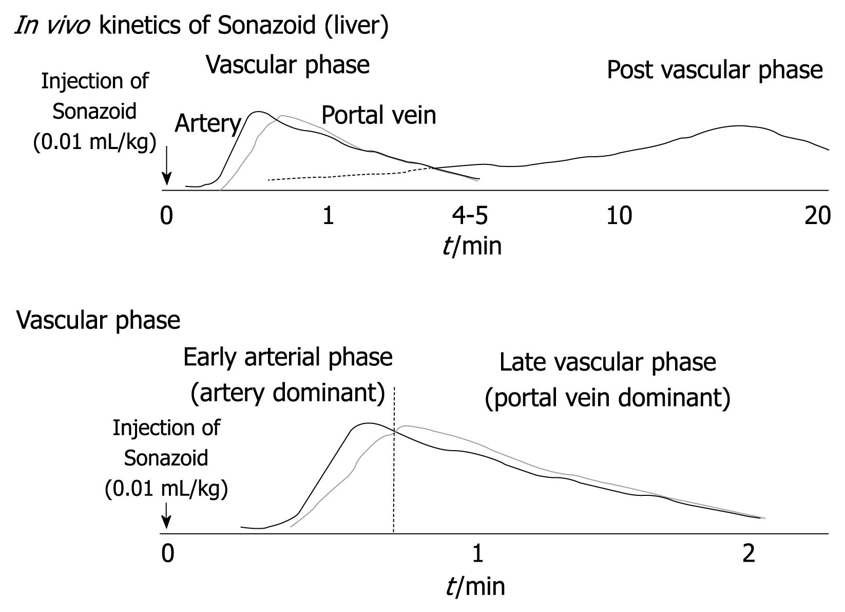
Figure 1 Kinetics of perfluorocarbon microbubbles (Sonazoid) in the liver.
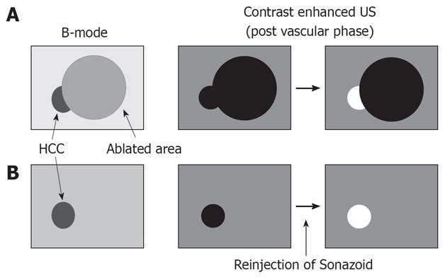
Figure 2 Indications for contrast-enhanced ultrasound (US) in radiofrequency (RF) ablation therapy.
A: Local tumor progression or residual hepatic malignancies. Contrast-enhanced US shows not only the tumor but also ablated areas as a defect image in the post vascular phase. However, viable tumor is enhanced by reinjection of Sonazoid; B: Hepatic malignancies with severe cirrhotic change. The true malignant tumor was sited among many large regenerated nodules in cirrhotic liver. On contrast-enhanced US, the tumor was distinguishable by reinjection of Sonazoid.
Figure 3 A 66-years-old man with 1.
0 cm hepatocellular carcinoma (HCC) (S3) with liver cirrhosis. A: Contrast harmonic US showed HCC nodule as a defect (arrow) in the post vascular phase after administration of Sonazoid; B: Enhancement (arrow) of HCC was obtained in the defect immediately after reinjection of Sonazoid; C: The defect (arrow) was later exhibited again and the RF electrode needle (arrowheads) was inserted.
- Citation: Minami Y, Kudo M. Contrast-enhanced harmonic ultrasound imaging in ablation therapy for primary hepatocellular carcinoma. World J Radiol 2009; 1(1): 86-91
- URL: https://www.wjgnet.com/1949-8470/full/v1/i1/86.htm
- DOI: https://dx.doi.org/10.4329/wjr.v1.i1.86


