Published online Aug 26, 2021. doi: 10.4330/wjc.v13.i8.254
Peer-review started: March 11, 2021
First decision: April 6, 2021
Revised: June 24, 2021
Accepted: July 26, 2021
Article in press: July 26, 2021
Published online: August 26, 2021
Processing time: 165 Days and 10.4 Hours
Infective endocarditis is one of the leading life-threatening infections around the world. With the exponential growth in the field of transcatheter interventions and advances in specialized surgical techniques, the number of prosthetic valves and cardiac implantable devices has significantly increased. This has led to a steep rise in the number of cases of prosthetic valve endocarditis (PVE) comprising up to 30% of all cases. Clinical guidelines rely on the use of the modified Duke criteria; however, the diagnostic sensitivity of the modified Duke criteria is reduced in the context of PVE. This is in part attributed to prosthesis related artifact which greatly affects the ability of echocardiography to detect early infective changes related to PVE in certain cases. There has been increasing recognition of the roles of complementary imaging modalities and updates in international society recommendations. Prompt diagnosis and treatment can prevent the devastating consequences of this condition. Imaging modalities such as cardiac computed tomography and 18-fluorodeoxyglucose positron emission tomography/computed tomography are diagnostic tools that provide a complementary role to echocardiography in aiding diagnosis, pre-operative planning, and treatment decision-making process in these challenging cases. Understanding the strengths and limitations of these adjuvant imaging modalities is crucial for the implementation of appropriate imaging modalities in clinical practice.
Core Tip: Prosthetic valve endocarditis comprises up to 30% of all cases of infective endocarditis with a reported in-hospital mortality of 14%-22% and 1-year mortality as high as 40%. Its prompt diagnosis, although often challenging, is of critical importance to prevent deleterious consequences for patients. Advances in the field of 3-dimen
- Citation: Lo Presti S, Elajami TK, Zmaili M, Reyaldeen R, Xu B. Multimodality imaging in the diagnosis and management of prosthetic valve endocarditis: A contemporary narrative review. World J Cardiol 2021; 13(8): 254-270
- URL: https://www.wjgnet.com/1949-8462/full/v13/i8/254.htm
- DOI: https://dx.doi.org/10.4330/wjc.v13.i8.254
Infective endocarditis (IE) is the third most common life-threatening infection in the world with a reported in-hospital mortality as high as 14%-22% and 1-year mortality of up to 40%[1]. The volume of prosthetic valve replacement procedures has dramatically increased over the last decades[2]. This has led to an increase in the incidence of prosthetic valve endocarditis (PVE), accounting for 20% to 30% of all cases of IE[2-4]. Traditionally, the diagnosis of IE is based on the modified Duke Criteria which relies on echocardiography[5]. Initially, transthoracic echocardiography (TTE) is utilized to assess for PVE; however, due to technical limitations and acoustic shadowing, transesophageal echocardiogram (TEE) is generally mandated when it is difficult to evaluate the prosthetic structures or the clinical suspicious remains high despite an apparently unremarkable TTE, according to the contemporary guidelines from the European Society of Cardiology (ESC) and American Heart Association/American College of Cardiology guidelines (AHA/ACC)[3,5,6]. In cases where TEE yields a negative result and clinical concern persists, guidelines recommend to either repeat the study in 3-7 days or to complement the evaluation with an alternative imaging modality such as 18-fluorodeoxyglucose photon emission tomography/computed tomography (PET/CT 18F-FDG) or cardiac computed tomography (CCT)[3,5,6]. Although TEE has generally good diagnostic performance, it is limited by prosthetic material-related artifacts, and certain complications of PVE, such as abscesses and pseudoaneurysms may be missed in some cases by TEE[7].
Patients with PVE are at a higher risk of developing complications and worse outcomes when compared with native valve endocarditis (NVE) patients, even when the causative organism are similar[3,8]. Therefore, it is of uttermost importance to accurately diagnose this condition and institute prompt treatment to ameliorate its deleterious consequences. There has been increased recognition of the pivotal role of multimodality imaging in the diagnosis and treatment of PVE[9,10]. The purpose of this narrative review is to focus on the diagnosis of PVE with a special emphasis on the emerging complementary use of multimodality imaging modalities.
The incidence of PVE ranges between 1% to 4% in the first year after surgery followed by approximately 1% per year thereafter[11]. The risk of developing PVE is higher within the first 5-years post-surgery (1.4% to 5.7%), and half of the patients with PVE develop a prosthetic valve abscess and pseudoaneurysm, which are associated with increased mortality of 30%-54%[7,12,13]. The reported incidence of PVE is heterogeneous, reflecting valve-related, patient, and geographical factors.
In a study from the Danish national registry of 18041 patients, the overall incidence of PVE was 69.8/10000 person years in patients undergoing surgical valve replacement[14]. When examined based on the anatomical location, the incidence was 65/10000 person years for mitral valve replacement (MVR), 70/10000 person years for aortic valve replacement (AVR), which increased to 89.4/10000 person years when both mitral and aortic valves were replaced[14]. Despite this difference, the cumulative incidence of PVE at 10-years was similar for both MVR and AVR (5.2%)[14].
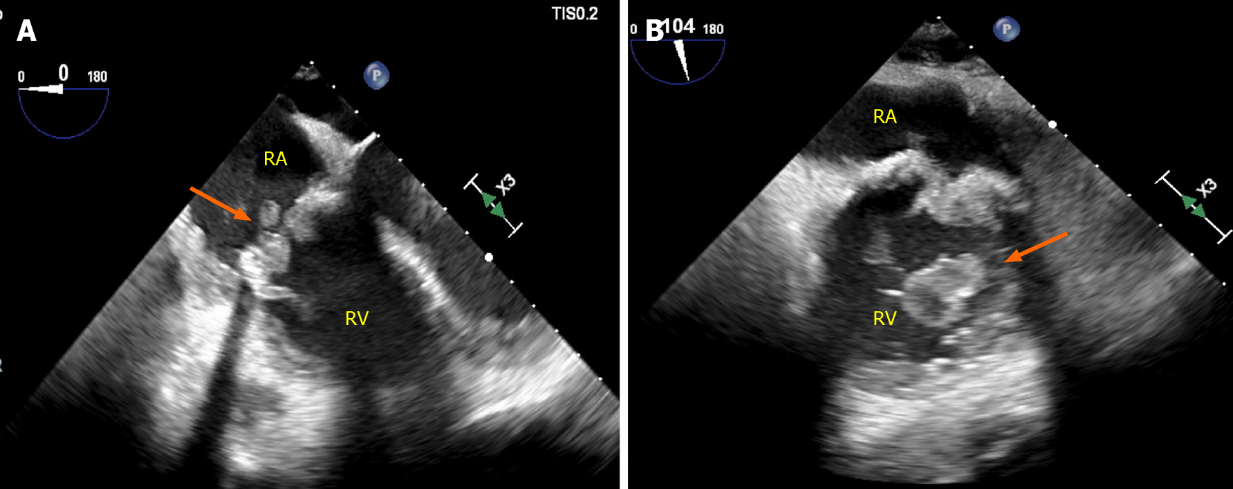
PVE comprises 11% of all cases of tricuspid valve endocarditis and 43% of all cases of pulmonary valve endocarditis (Figure 1)[15]. In a cohort of congenital heart disease patients, 924 surgical pulmonic valve replacement were performed with 19 (2%) cases attributed to PVE, corresponding to an incidence of 333/100000 person years[16]. A large single-center cohort of 2124 adult patients (median age 41.5 years) with IE reported 24 cases of pulmonary valve endocarditis, of which 54.2% of cases occurred in the context of prosthetic valves[17].
PVE is also a feared complication in transcatheter aortic valve replacement (TAVR)[18]. In a meta-analysis by Ando et al[19] in 3761 patients undergoing percutaneous or surgical AVR (SAVR), the overall incidence of PVE was not significantly different between TAVR and SAVR at 1, 2 and 3.4 years follow-up[19]. Over this period of time, there was a trend towards a higher incidence of PVE in the TAVR group (0.86% to 2%) compared to the SAVR group (0.73% to 1.3%), and this was enhanced in patients with intermediate surgical risk [2.3% vs 1.2%; odds ratio (OR) = 1.92, 95% confidence intervals (CI): 0.99 to 3.72, P = 0.05]. In the Nordic Aortic Valve Intervention (NOTION) trial, 280 Low-surgical risk patients with severe aortic stenosis were randomized to TAVR (self-expanding CoreValve) or SAVR (stented bioprosthesis)[20]. This trial showed a non-significant difference in the 5 year-cumulative incidence of PVE between these two approaches (6.2% for TAVR vs 4.4% SAVR)[20]. Similar results were reported by Summer et al[21], in a pooled cohort of all patients from PARTNER I and PARTNER II trials (8530 patients, 107 cases of PVE), where the incidence over time of PVE was similar for TAVR [5.21 PVE per 1000 person-years (95%CI: 4.26–6.38)] and for SAVR [4.10 per 1000 person-years (95%CI: 2.33–7.22); incident rate ratio, 1.27 (95%CI: 0.70–2.32); P = 0.44][21]. Ando et al[19] also reported a subgroup analysis in TAVR patients which demonstrated comparable risk between balloon-expandable valves (BEV) and self-expandable valves (SEV)[19]. Similar findings were described by Regueiro et al[22] in a cohort of 6363 patients undergoing TAVR, where the incidence of PVE at 1-year did not significantly differ (0.95% SEV vs 1.25% BEV; P = 0.33)[22]. When other complications were analyzed, the rate of systemic stroke and embolism was higher in patients with BEV (8.7% vs 20.0% adjusted OR = 2.46, 95%CI: 1.04–5.82, P = 0.04)[22].
Furthermore, percutaneous edge to edge mitral valve repair is an increasingly relevant transcatheter intervention, where post clip implantation endocarditis has been described only in case reports, remaining an extremely rare presentation[23].
In terms of the type of valve prosthesis, the Danish National Registry demonstrated in 18041 patients undergoing left-sided valve replacement that the use of bioprosthetic valves was associated with an increased risk for prosthetic infection in patient undergoing either MVR (HR = 1.91, 95%CI: 1.08–3.37) or AVR (HR = 1.70, 95%CI: 1.35–2.15) over 10 years follow-up[14]. These results were similar to those reported by Brennan et al[24] in a large cohort of patients undergoing SAVR (bioprosthetic = 24410; and mechanical = 14789) followed up for 12 years where the risk of PVE was higher in those patients undergoing bioprosthetic valve replacement (HR = 1.60; 95%CI: 1.31–1.94). However, a limitation of these studies was their retrospective nature [14,24]. Conflicting evidence arises from 3 randomized clinical trials, comprising a total of 40207 patients that underwent left sided valve replacement, which showed no sig
There is conflicting data regarding the age group most susceptible to develop PVE. In the Grupo de Apoyo al Manejo de la Endocarditis infecciosa en España (GAMES) Database registry, which included 3120 patients with IE, patients 65-79 years old (elderly) had a significantly higher incidence of PVE compared with those < 65 years (young) and ≥ 80 years old (octogenarian) (37.3% vs 24.4% and 26.3%, respectively, P < 0.001)[28]. This observation was also demonstrated by López et al[29], studying a cohort of 600 Left-sided IE patients 40% of whom had PVE, showing a similar age distribution[29]. In contrast, a smaller observational study of 72 patients with IE by Menchi-Elanzi et al[30] demonstrated that elderly patients (65-79 years old) had a significantly lower prevalence of PVE compared to the young and octogenarians[30].
In patients with PVE, there is male predominance with 3:1 ratio across various studies[14,28-30]. This ratio changes towards 1:1 in the octogenarian group[28-30]. Interestingly, in an observational study of 621 patients with left-sided IE, mitral mechanical valve PVE was more common in women than in men[31]. The mechanism behind these findings remain unclear, and further studies are required to examine the age and gender influence on PVE.
In an observational, prospective multicenter cohort of 2670 patients from 28 countries with IE, 556 patients (20.1%) had PVE[1].The highest percentage of PVE cases was in Southern Europe, Middle East and South Africa (26.10%), and it was the lowest in South America (11.9%)[2]. In the United States, the incidence of PVE was 20.9%, with the highest source corresponding to health care associated infections (44.8%), followed by intravascular device-related infection (27.6%), non-nosocomial heath care-associa
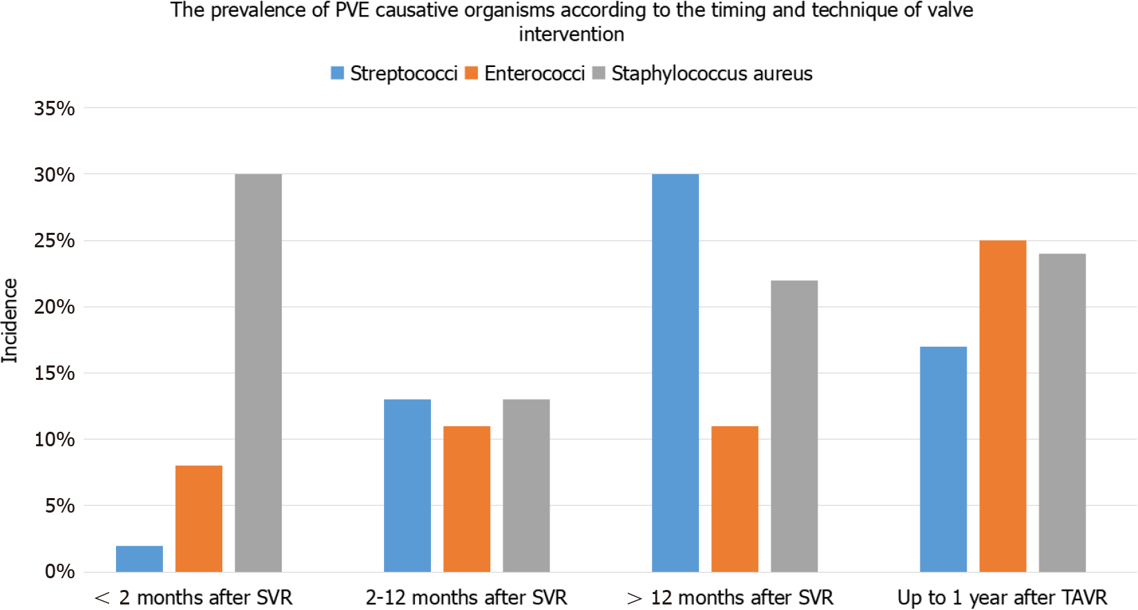
The prevalence of the most common causative organisms causing PVE according to the timing and technique of valve surgery are shown in Figure 2. Within 60 days from surgical valve replacement, the most common causative organism of PVE is Staphylococcus Aureus (30%) followed by Streptococcus species (28%). Between 2 to 12 months after surgery, coagulase-negative Staphylococci are the most common organism (36%), and after one year, Streptococci predominantly viridians group, are the leading cause[2,32-35]. After TAVR, Enterococci and Staphylococcus Aureus (25% and 16%-24%, respectively) are the predominant organisms (Figure 2)[36,37].
The clinical diagnosis of PVE is challenging as patients often manifest non-specific symptoms, such as fever, weakness and poor appetite in the early post-operative period[38]. Therefore, the presence of a new murmur, new or worsening congestive heart failure (CHF), conduction abnormalities and stroke should all raise suspicion for PVE[38]. Data from the International Collaboration on Endocarditis (ICE) Prospective Cohort Study (PCS) reported the occurrence of CHF in 32.9%, intra-cardiac abscess in 29.7%, stroke in 18.2% and other systemic embolization in 14.9%, among 556 patients with PVE[2]. When comparing NVE and PVE, both had a similar incidence of CHF, stroke and persistent bacteremia; however, the incidence of systemic embolization was lower in PVE[2].
From the TAVR international registry consisting of 245 patients who developed PVE after TAVR (BEV and SEV), the most common initial symptom was fever (approximately 80%), followed by CHF (approximately 40%), and cutaneous manifestations (approximately 3%) with a median time to onset after TAVR of 5.3-5.5 months[22]. In this study, stroke was significantly higher with BEV compared to SEV (24.6% vs 7.8%, P < 0.01)[22]. The lower rate of cutaneous manifestations in PVE, such as Osler’s nodes, Janeway lesions and Roth’s spots could be attributed to a more acute course of the disease in PVE, compared to a more protracted course commonly seen in NVE[38]. In Reguiero’s cohort, patients with PVE following TAVR had no significant difference in mortality between BEV and SEV (37% vs 36%, respectively)[22].
In terms of mortality, Wang et al[2], described in-hospital mortality was significantly higher in the PVE group (127/556 patients) compared to NVE (310/1895 patients) (23% vs 16%, P < 0.001). After multivariate analysis, the key drivers of increased mortality were CHF (OR = 2.33, 95%CI: 1.62-3.34), intracardiac abscess (OR = 1.86, 95%CI: 1.10-3.15), and stroke (OR = 2.25, 95%CI: 1.25-4.03). Østergaard et al[14] in a cohort of 18,041 undergoing left sided valve replacement (AVR 88.8%, MVR 9.7%, and both 1.5%) demonstrated that PVE in AVR patients was associated with higher mortality than in MVR at 10 years (44% vs 39%, P < 0.01)[14]. Moreover, they also divided these results according to the prosthesis type showing a significantly higher mortality with bioprosthetic compared to mechanical valve in both AVR and MVR at 10-years. However, when both groups were matched, there was no significant diffe
Although acoustic shadowing and reverberation artifacts from prosthetic material hamper the imaging resolution, echocardiography remains the forefront diagnostic modality in suspected PVE[5]. Wide availability, low cost, rapid acquisition and interpretation, and a lack of radiation are some of the important qualities that make echocardiography the first-line imaging modality[6]. The echocardiographic exami
The sensitivity of TTE in PVE ranges from 17% to 36%; in comparison, it increases to 82% to 96% with TEE, suggesting the importance of TEE for better assessment of all cases of suspected PVE[39,40]. Despite the enhanced temporal and spatial resolution of multiplanar TEE, its ability to identify prosthetic valve abnormalities can be cha
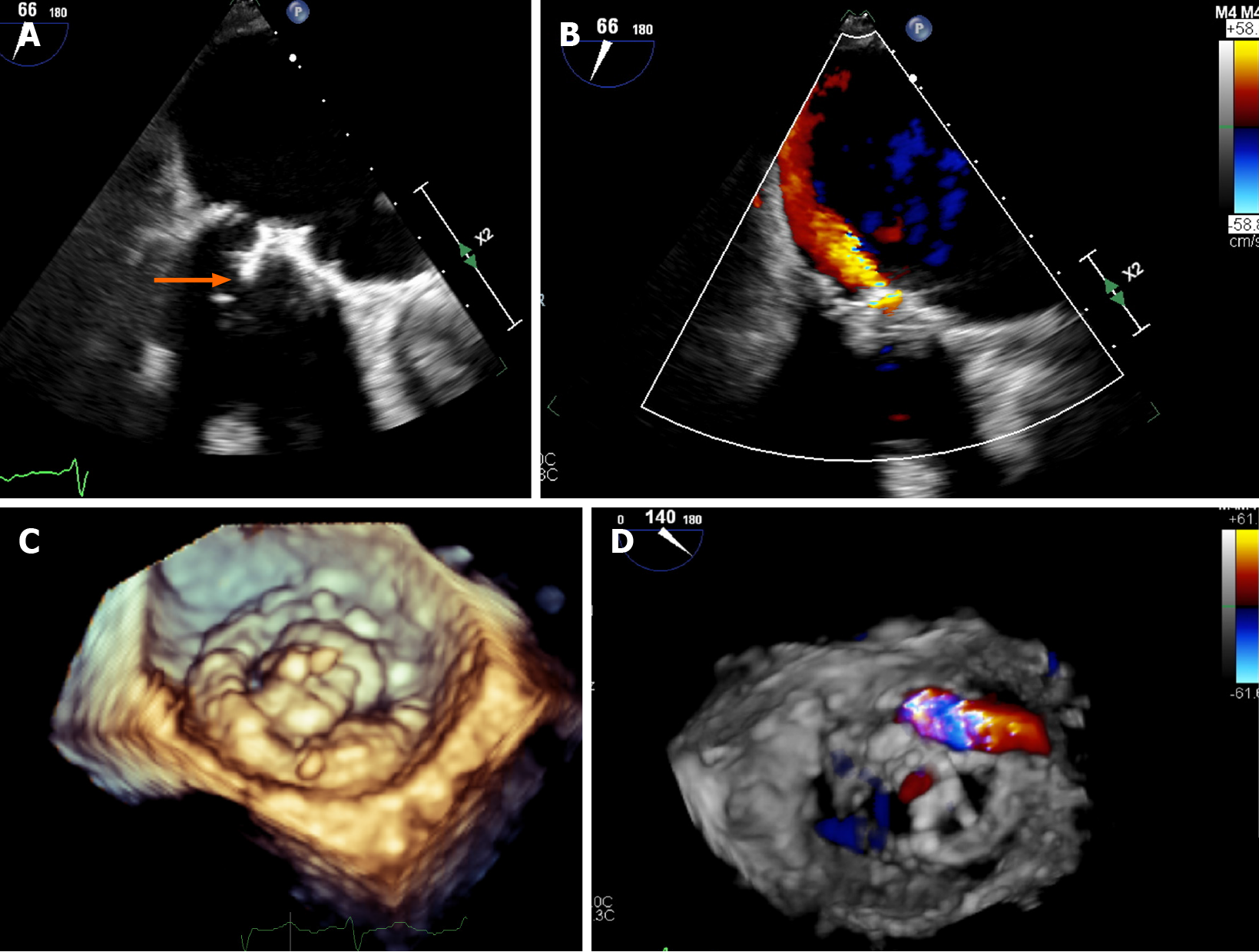
Three-dimensional (3D) echocardiography is a complementary modality which provides valuable information regarding the anatomy of the prosthetic valves and adjacent structures from different angles. Novel 3D-rendering software aid in the characterization of the vegetation size and location, destructive changes, perforations, abscess characterization, prosthetic dehiscence and associated regurgitant jets[42-44]. Chahine et al[45] described in 242 patients, an improved sensitivity over the recent decade for the detection of PVE (70.8% vs 93.7%, P = 0.009) with contemporary TEE technology including an increased use of 3D imaging (Figure 4)[45].
Echocardiography has been shown to predict outcomes in PVE. Wang et al[46] studied 115 patients with surgically proven IE (52% with bioprosthetic valves; 15.5% with metallic valves) and recognized that abscess or pseudoaneurysm detected by TEE were independently associated with increased in-hospital mortality and morbidity [OR: 3.66 (95%CI: 1.76–7.59); P = 0.001][46].
In cases where the clinical suspicion remains high despite an initial negative result, short-term interval follow-up is a strategy that can enhance imaging sensitivity at the expense of prolonging the time to diagnosis. This can usually be performed 3-7 days following the initial evaluation[3]. In contrast to this “watch and wait approach”, adjuvant imaging modalities play a complementary role in the diagnosis of PVE, potentially expediting patient care[7].
CCT has become an increasingly important imaging tool for the diagnosis and pre-operative planning of patients with PVE. CCT offers a number of technical advantages over echocardiography including higher spatial resolution and imaging window independence[47]. CCT has demonstrated similar diagnostic yield for the detection of perivalvular complication[7]. Feutcher et al[47] compared CCT with TEE in 37 patients with IE, 6 of whom had PVE. The study showed that CCT had an excellent correlation with TEE in determining vegetation size (vegetation size by TEE 7.6 ± 5.6 mm) (r = 0.95; P < 0.001). In addition, vegetation mobility was accurately diagnosed by CCT in 96% of the patients, and both modalities had similar detection rates for abscesses and pseudoaneurysms with the caveat that CCT provided more detailed anatomical lo
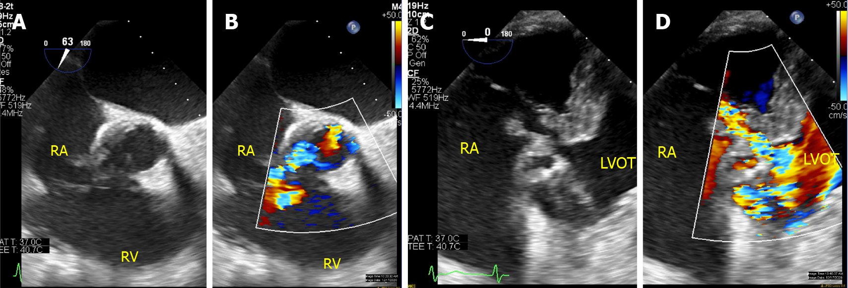
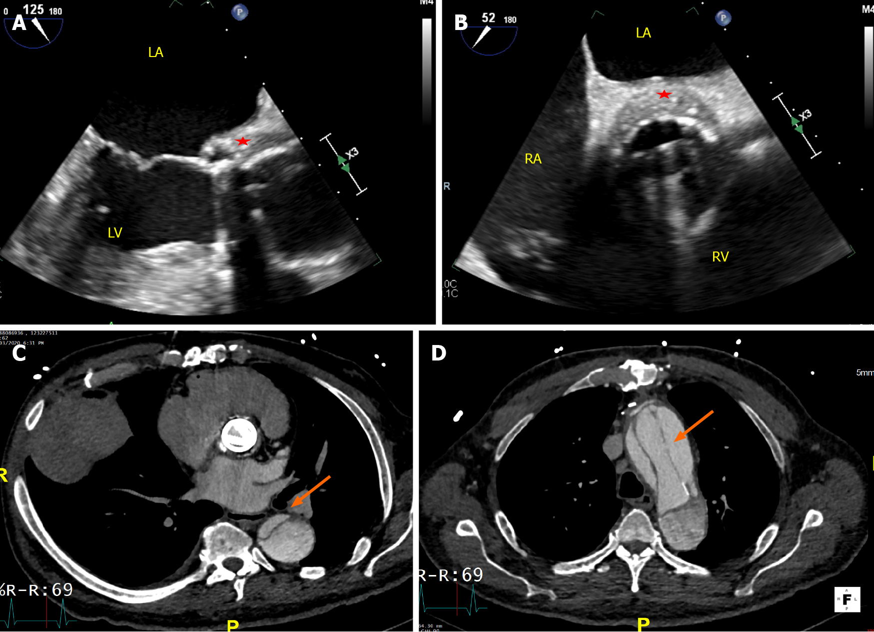
CCT has also proven to be useful in surgical planning by providing additional diagnostic information regarding the anatomy of coronary arteries and aorta, and degree of calcification[3,51]. The major advantage of CCT over coronary angiography, is the ability to demonstrate coronaries and bypass graft patency non-invasively, avoiding risk of vegetation embolization during catheter manipulation[47]. It may also be valuable in the urgent evaluation of hemodynamically unstable patients who are unable to undergo TEE (Figures 5 and 6)[52].
A potential weakness of CCT is its relatively low temporal resolution, resulting in decreased sensitivity for the detection of small vegetations (< 4 mm) and leaflet per
| Modality | Strengths | Weaknesses | Sensitivity | Specificity |
| Echocardiography | Available, convenient, no radiation exposure, hemodynamic data, high temporal resolution. | Operator- and imaging window-dependent, affected by prosthesis-related artifacts. | TTE: 17%–36%; TEE: 82%–96% | TTE: 86%[39,40]; TEE: 94%[39,40,45] |
| Cardiac CT | Spatial resolution, defining paravalvular complications, delineating coronary-aorta anatomy, preoperative planning. | Radiation exposure, contrast exposure limits use in advanced CKD. | 88%–97% | 95%[47,48,50] |
| Cardiac MRI | Characterizing paravalvular complications, depicts inflammatory changes, assess the degree of intra-cardiac shunting. | Limited data, lower spatial resolution, incompatibility with some cardiac devices. Limited clinical applicability. | Limited data | Limited data |
| 18F-FDG PET/CT | Excellent diagnostic role in PVE, detection of metastatic infection foci. | Availability, cost, requires special pre-test preparation, expertise, radiation exposure. | 73%–97% | 80%–94%[60-62] |
The roles of cardiac magnetic resonance imaging (MRI) in IE, and more specifically in PVE, are currently limited, and less well defined. In general, this imaging modality offers several unique advantages such as improved 3D-visualization of cardiac structures compared to TEE, the ability to identify inflammatory changes in the myocardium and pericardium via delayed enhancement imaging, differentiation of vegetations from intracardiac masses, ability to diagnose infiltrative cardiomyopathies, accurate quantification of regurgitant valvular lesions and the ability to be used in patients unable to receive iodine-based contrast[54,55]. However, the role of MRI for evaluation of infective changes, especially in PVE is limited. Some of the factors that account for its limitations include incompatibility with some implantable cardiac devices, reduced availability and significant artifacts caused by metallic leaflets[53,54].
As part of the pre-operative work up, MRI does not provide as accurate information regarding the anatomy of the chest wall and its proximity to cardiac structures as does CCT, and thus is not usually favored for this purpose[56]. Nevertheless, MRI of the brain is recommended in pre-operative patients who have neurologic deficits and may also be reasonable in high-risk left-sided IE to screen for subclinical embolic events[3,56]. The ability of brain MRI to detect subclinical cerebral lesions, which may be found in up to 70% of patients who are neurologically intact clinically, has substantial clinical implications, as presence of systemic embolization represents one minor Duke criterion[5]. This in turn may allow earlier diagnosis and the implementation of thera
Hybrid modalities such as leucocyte scintigraphy and 18F-FDG PET/CT have also been recognized as important complementary diagnostic imaging modalities. 18F-FDG PET/CT relies on the administration 18F-FDG radioisotope, which is taken up by active inflammatory cells at the site of the infection. On the other hand, leucocyte scintigraphy isolates and labels granulocytes with 99mTc that can be localized and quantified at a specific acquisition point in time[3,59]. The steps in preparation of leucocyte scintigraphy involving the drawing and reinjection of leucocytes, makes the utilization of 18F-FDG radioisotope more favorable in clinical practice[1].
The modified Duke criteria only considers echocardiography as the diagnostic imaging modality for IE. The AHA guidelines, despite acknowledging the usefulness of PET/CT for the detection of extracardiac complications, have not yet recommended its routine use for diagnosis[5]. In contrast, in the latest iteration of the ESC guidelines for the management of IE, the presence of abnormal activity of 18F-FDG PET/CT or leucocyte scintigraphy SPECT/CT (> 3 months after implant) around the perivalvular region of a prosthetic valve was upgraded as a major imaging criterion for diagnosis of IE[3]. As a result, Saby et al[60] reported an improvement of the modified Duke criteria sensitivity from 70% to 97% without trading-off its specificity, with the use of PET/CT [60]. Although similar findings in terms of sensitivity were found by Philip et al[61] in 115 patients with PVE (91 definite cases and 24 rejected cases) where the sensitivity increased from 57.1% to 83.5%; there was a decrease in specificity from 95.8% to 70.8%, with an overall improvement in accuracy from 65.2% to 80.9%. Wang et al[62] also reported in 333 patients with PVE an enhanced sensitivity of 86%, however, the sensitivity of the test decreased to 72% in the presence of cardiac implantable elec
18F-FDG PET/CT can be utilized early in the evaluation of suspected PVE, especially if microbiologic cultures and echocardiographic imaging are unrevealing[63]. 18F-FDG PET/CT is a useful complimentary imaging modality for the diagnosis of IE that has demonstrated improving performance over time, especially in challenging cases of PVE and CIED related infections[62,64]. It has also been suggested to have a potential role in monitoring the response to antibiotic therapy[63,65]. The enhanced diagnosis of PVE with PET/CT has important clinical implications, helping to re-classify up to 90% of the “possible IE” cases by modified Duke Criteria, and providing a conclusive diagnosis (definite/rejected) in 95% of the cases[66]. It has also significantly altered the treatment plan in up to 35% of the cases by virtue of antibiotic treatment prolongation (27.5%), surgical referral (15%) and prevention of unnecessary device extraction (17.7%)[67]. This is attributed in part to its ability to detect extracardiac foci of in
Cautious interpretation of 18F-FDG PET/CT results must be entertained, especially in the early postsurgical period during the first 3 months. Following the implantation of a prosthetic valve, an inflammatory response to the foreign body occurs, which is reactive in nature without necessarily implying the presence of infection[69]. Other causes of false positive results include: soft atherosclerotic plaques and active thrombi, cardiac tumors (whether primary or metastatic), and inflammatory conditions such as vasculitis and myocarditis[70,71]. Rouzet et al[59] described in a cohort of 39 patients with prosthetic valves and absence of clinical infection that approximately half of these patients will continue to have a homogenous uptake on 18F-FDG PET/CT in the perivalvular area that may persist years after surgery, and therefore should not be confused with infective changes[59]. On the other hand, false negative may still occur in the presence of small vegetations (< 5 mm), recent antibiotic administration, metastatic brain lesions and high glucose states[3,72]. Although contemporary data reported equipment availability in 70.3% of European centers and 56.3% non-ESC centers, the availability, cost, and expertise needed with this imaging modality impose additional limitations on its employment in routine clinical practice[4,73].
Real world data from the ESC-EORP EURO-ENDO (European infective endocar
In this section, a brief overview of the general management principles will be discu
Antibiotic therapy should be initiated in all cases of PVE with consultation of an infectious disease specialist for guiding the antibiotic choice[3,5,38,56]. Three sets of blood cultures separated by 30-60 minutes should be obtained before antibiotic initiation and at least every 24-48 h until the blood culture is negative[3,5,38,56]. All patients should be monitored for side effects of antibiotics, clinical response and symptoms/signs that suggest PVE complications. In the latter case, an echocardiogram should be repeated. The treatment duration is generally 6 week starting from the first negative blood culture, and it can be extended for an additional 6 week if surgical specimens demonstrate a positive gram stain and culture or positive polymerase chain reaction. Antibiotic therapy should subsequently be tailored according to the culture results[3,5,38,56].
The diagnosis of PVE remains challenging due to its often non-specific clinical presentations and prosthesis-related artifacts that impair the optimal visualization of cardiac structures by echocardiography. Echocardiography continues to be the first-line imaging modality in suspected cases of PVE due to is wide availability, low cost, rapid interpretation, and safety. TEE is mandated in most PVE cases due to the reduced sensitivity of TTE in this context. The consequences of missing prosthesis-related infections are serious, and therefore, evaluation of PVE requires the optimal complementary use of imaging modalities to achieve the best outcomes. Adjuvant imaging modalities, particularly CCT and 18F-FDG PET/CT have important niche roles. These imaging modalities improve the ability to accurately and timely diagnose PVE, contribute to the pre-operative planning of appropriate patients, and guide decision-making for therapies.
Manuscript source: Invited manuscript
Specialty type: Cardiac and cardiovascular systems
Country/Territory of origin: United States
Peer-review report’s scientific quality classification
Grade A (Excellent): A
Grade B (Very good): B
Grade C (Good): 0
Grade D (Fair): D, D
Grade E (Poor): 0
P-Reviewer: Barik R, Gulel O S-Editor: Ma YJ L-Editor: A P-Editor: Yuan YY
| 1. | Gomes A, Glaudemans AWJM, Touw DJ, van Melle JP, Willems TP, Maass AH, Natour E, Prakken NHJ, Borra RJH, van Geel PP, Slart RHJA, van Assen S, Sinha B. Diagnostic value of imaging in infective endocarditis: a systematic review. Lancet Infect Dis. 2017;17:e1-e14. [RCA] [PubMed] [DOI] [Full Text] [Cited by in Crossref: 142] [Cited by in RCA: 190] [Article Influence: 21.1] [Reference Citation Analysis (0)] |
| 2. | Wang A, Athan E, Pappas PA, Fowler VG Jr, Olaison L, Paré C, Almirante B, Muñoz P, Rizzi M, Naber C, Logar M, Tattevin P, Iarussi DL, Selton-Suty C, Jones SB, Casabé J, Morris A, Corey GR, Cabell CH; International Collaboration on Endocarditis-Prospective Cohort Study Investigators. Contemporary clinical profile and outcome of prosthetic valve endocarditis. JAMA. 2007;297:1354-1361. [RCA] [PubMed] [DOI] [Full Text] [Cited by in Crossref: 447] [Cited by in RCA: 453] [Article Influence: 25.2] [Reference Citation Analysis (0)] |
| 3. | Habib G, Lancellotti P, Antunes MJ, Bongiorni MG, Casalta JP, Del Zotti F, Dulgheru R, El Khoury G, Erba PA, Iung B, Miro JM, Mulder BJ, Plonska-Gosciniak E, Price S, Roos-Hesselink J, Snygg-Martin U, Thuny F, Tornos Mas P, Vilacosta I, Zamorano JL; ESC Scientific Document Group . 2015 ESC Guidelines for the management of infective endocarditis: The Task Force for the Management of Infective Endocarditis of the European Society of Cardiology (ESC). Endorsed by: European Association for Cardio-Thoracic Surgery (EACTS), the European Association of Nuclear Medicine (EANM). Eur Heart J. 2015;36:3075-3128. [RCA] [PubMed] [DOI] [Full Text] [Cited by in Crossref: 2661] [Cited by in RCA: 3342] [Article Influence: 334.2] [Reference Citation Analysis (0)] |
| 4. | Habib G, Erba PA, Iung B, Donal E, Cosyns B, Laroche C, Popescu BA, Prendergast B, Tornos P, Sadeghpour A, Oliver L, Vaskelyte JJ, Sow R, Axler O, Maggioni AP, Lancellotti P; EURO-ENDO Investigators. Clinical presentation, aetiology and outcome of infective endocarditis. Results of the ESC-EORP EURO-ENDO (European infective endocarditis) registry: a prospective cohort study. Eur Heart J. 2019;40:3222-3232. [RCA] [PubMed] [DOI] [Full Text] [Cited by in Crossref: 219] [Cited by in RCA: 485] [Article Influence: 97.0] [Reference Citation Analysis (0)] |
| 5. | Baddour LM, Wilson WR, Bayer AS, Fowler VG Jr, Tleyjeh IM, Rybak MJ, Barsic B, Lockhart PB, Gewitz MH, Levison ME, Bolger AF, Steckelberg JM, Baltimore RS, Fink AM, O'Gara P, Taubert KA; American Heart Association Committee on Rheumatic Fever; Endocarditis, and Kawasaki Disease of the Council on Cardiovascular Disease in the Young, Council on Clinical Cardiology, Council on Cardiovascular Surgery and Anesthesia, and Stroke Council. Infective Endocarditis in Adults: Diagnosis, Antimicrobial Therapy, and Management of Complications: A Scientific Statement for Healthcare Professionals From the American Heart Association. Circulation. 2015;132:1435-1486. [RCA] [PubMed] [DOI] [Full Text] [Cited by in Crossref: 1569] [Cited by in RCA: 2091] [Article Influence: 209.1] [Reference Citation Analysis (1)] |
| 6. | Haq IU, Haq I, Griffin B, Xu B. Imaging to evaluate suspected infective endocarditis. Cleve Clin J Med. 2021;88:163-172. [RCA] [PubMed] [DOI] [Full Text] [Cited by in Crossref: 21] [Cited by in RCA: 16] [Article Influence: 4.0] [Reference Citation Analysis (0)] |
| 7. | Habets J, Tanis W, van Herwerden LA, van den Brink RB, Mali WP, de Mol BA, Chamuleau SA, Budde RP. Cardiac computed tomography angiography results in diagnostic and therapeutic change in prosthetic heart valve endocarditis. Int J Cardiovasc Imaging. 2014;30:377-387. [RCA] [PubMed] [DOI] [Full Text] [Cited by in Crossref: 59] [Cited by in RCA: 70] [Article Influence: 5.8] [Reference Citation Analysis (0)] |
| 8. | Lalani T, Chu VH, Park LP, Cecchi E, Corey GR, Durante-Mangoni E, Fowler VG Jr, Gordon D, Grossi P, Hannan M, Hoen B, Muñoz P, Rizk H, Kanj SS, Selton-Suty C, Sexton DJ, Spelman D, Ravasio V, Tripodi MF, Wang A; International Collaboration on Endocarditis–Prospective Cohort Study Investigators. In-hospital and 1-year mortality in patients undergoing early surgery for prosthetic valve endocarditis. JAMA Intern Med. 2013;173:1495-1504. [RCA] [PubMed] [DOI] [Full Text] [Cited by in Crossref: 164] [Cited by in RCA: 202] [Article Influence: 16.8] [Reference Citation Analysis (0)] |
| 9. | Habib G, Derumeaux G, Avierinos JF, Casalta JP, Jamal F, Volot F, Garcia M, Lefevre J, Biou F, Maximovitch-Rodaminoff A, Fournier PE, Ambrosi P, Velut JG, Cribier A, Harle JR, Weiller PJ, Raoult D, Luccioni R. Value and limitations of the Duke criteria for the diagnosis of infective endocarditis. J Am Coll Cardiol. 1999;33:2023-2029. [RCA] [PubMed] [DOI] [Full Text] [Cited by in Crossref: 206] [Cited by in RCA: 205] [Article Influence: 7.9] [Reference Citation Analysis (0)] |
| 10. | Habib G, Thuny F, Avierinos JF. Prosthetic valve endocarditis: current approach and therapeutic options. Prog Cardiovasc Dis. 2008;50:274-281. [RCA] [PubMed] [DOI] [Full Text] [Cited by in Crossref: 117] [Cited by in RCA: 116] [Article Influence: 6.8] [Reference Citation Analysis (0)] |
| 11. | Dhawan VK. Infective endocarditis in elderly patients. Clin Infect Dis. 2002;34:806-812. [RCA] [PubMed] [DOI] [Full Text] [Cited by in Crossref: 61] [Cited by in RCA: 50] [Article Influence: 2.2] [Reference Citation Analysis (0)] |
| 12. | Daniel WG, Mügge A, Martin RP, Lindert O, Hausmann D, Nonnast-Daniel B, Laas J, Lichtlen PR. Improvement in the diagnosis of abscesses associated with endocarditis by transesophageal echocardiography. N Engl J Med. 1991;324:795-800. [RCA] [PubMed] [DOI] [Full Text] [Cited by in Crossref: 516] [Cited by in RCA: 446] [Article Influence: 13.1] [Reference Citation Analysis (0)] |
| 13. | Hill EE, Herijgers P, Claus P, Vanderschueren S, Peetermans WE, Herregods MC. Abscess in infective endocarditis: the value of transesophageal echocardiography and outcome: a 5-year study. Am Heart J. 2007;154:923-928. [RCA] [PubMed] [DOI] [Full Text] [Cited by in Crossref: 133] [Cited by in RCA: 138] [Article Influence: 7.7] [Reference Citation Analysis (0)] |
| 14. | Østergaard L, Valeur N, Ihlemann N, Smerup MH, Bundgaard H, Gislason G, Torp-Pedersen C, Bruun NE, Køber L, Fosbøl EL. Incidence and factors associated with infective endocarditis in patients undergoing left-sided heart valve replacement. Eur Heart J. 2018;39:2668-2675. [RCA] [PubMed] [DOI] [Full Text] [Cited by in Crossref: 34] [Cited by in RCA: 56] [Article Influence: 9.3] [Reference Citation Analysis (0)] |
| 15. | Akinosoglou K, Apostolakis E, Koutsogiannis N, Leivaditis V, Gogos CA. Right-sided infective endocarditis: surgical management. Eur J Cardiothorac Surg. 2012;42:470-479. [RCA] [PubMed] [DOI] [Full Text] [Cited by in Crossref: 80] [Cited by in RCA: 86] [Article Influence: 6.6] [Reference Citation Analysis (0)] |
| 16. | Robichaud B, Hill G, Cohen S, Woods R, Earing M, Frommelt P, Ginde S. Bioprosthetic pulmonary valve endocarditis: Incidence, risk factors, and clinical outcomes. Congenit Heart Dis. 2018;13:734-739. [RCA] [PubMed] [DOI] [Full Text] [Cited by in Crossref: 9] [Cited by in RCA: 12] [Article Influence: 1.7] [Reference Citation Analysis (0)] |
| 17. | Isaza N, Shrestha NK, Gordon S, Pettersson GB, Unai S, Vega Brizneda M, Witten JC, Griffin BP, Xu B. Contemporary Outcomes of Pulmonary Valve Endocarditis: A 16-Year Single Centre Experience. Heart Lung Circ. 2020;29:1799-1807. [RCA] [PubMed] [DOI] [Full Text] [Cited by in Crossref: 11] [Cited by in RCA: 13] [Article Influence: 2.6] [Reference Citation Analysis (0)] |
| 18. | Kaur S, Misbah Rameez R, Jaber W, Griffin BP, Xu B. Transcatheter Aortic Valve Replacement Associated Infective Endocarditis: A Clinical Update. Structural Heart. 2020;4:152-158. [RCA] [DOI] [Full Text] [Cited by in Crossref: 2] [Cited by in RCA: 4] [Article Influence: 0.8] [Reference Citation Analysis (0)] |
| 19. | Ando T, Ashraf S, Villablanca PA, Telila TA, Takagi H, Grines CL, Afonso L, Briasoulis A. Meta-Analysis Comparing the Incidence of Infective Endocarditis Following Transcatheter Aortic Valve Implantation Versus Surgical Aortic Valve Replacement. Am J Cardiol. 2019;123:827-832. [RCA] [PubMed] [DOI] [Full Text] [Cited by in Crossref: 32] [Cited by in RCA: 53] [Article Influence: 8.8] [Reference Citation Analysis (0)] |
| 20. | Thyregod HGH, Ihlemann N, Jørgensen TH, Nissen H, Kjeldsen BJ, Petursson P, Chang Y, Franzen OW, Engstrøm T, Clemmensen P, Hansen PB, Andersen LW, Steinbrüchel DA, Olsen PS, Søndergaard L. Five-Year Clinical and Echocardiographic Outcomes from the Nordic Aortic Valve Intervention (NOTION) Randomized Clinical Trial in Lower Surgical Risk Patients. Circulation. 2019;. [RCA] [PubMed] [DOI] [Full Text] [Cited by in Crossref: 191] [Cited by in RCA: 245] [Article Influence: 40.8] [Reference Citation Analysis (0)] |
| 21. | Summers MR, Leon MB, Smith CR, Kodali SK, Thourani VH, Herrmann HC, Makkar RR, Pibarot P, Webb JG, Leipsic J, Alu MC, Crowley A, Hahn RT, Kapadia SR, Tuzcu EM, Svensson L, Cremer PC, Jaber WA. Prosthetic Valve Endocarditis After TAVR and SAVR: Insights From the PARTNER Trials. Circulation. 2019;140:1984-1994. [RCA] [PubMed] [DOI] [Full Text] [Cited by in Crossref: 53] [Cited by in RCA: 79] [Article Influence: 13.2] [Reference Citation Analysis (0)] |
| 22. | Regueiro A, Linke A, Latib A, Ihlemann N, Urena M, Walther T, Husser O, Herrmann C, Nombela-Franco L, Cheema A, Le Breton H, Stortecky S, Kapadia S, Bartorelli L, Sinning JM, Amat-Santos I, Munoz-Garcia J, Lerakis S, Gutíerrez-Ibanes E, Abdel-Wahab M, Tchetche D, Testa L, Eltchaninoff H, Livi U, Castillo JC, Jilaihawi H, Webb G, Barbanti M, Kodali S, de Brito Jr S, Ribeiro B, Miceli A, Fiorina C, Actis Dato GM, Rosato F, Serra V, Masson JB, Wijeysundera C, Mangione A, Ferreira MC, Lima C, Carvalho A, Abizaid A, Marino A, Esteves V, Andrea C. M., Messika-Zeitoun D, Himbert D, Kim WK, Pellegrini C, Auffret V, Nietlispach F, Pilgrim T, Durand E, Lisko J, Makkar R, Lemos P, Leon B, Puri R, San Roman A, Vahanian A, Søndergaard L, Mangner N, Rodés-Cabau J. Infective Endocarditis Following Transcatheter Aortic Valve Replacement: Comparison of Balloon- Versus Self-Expandable Valves. Circ Cardiovasc Interv. 2019;12:e007938. [RCA] [PubMed] [DOI] [Full Text] [Cited by in Crossref: 22] [Cited by in RCA: 40] [Article Influence: 6.7] [Reference Citation Analysis (0)] |
| 23. | Boeder NF, Dörr O, Rixe J, Weipert K, Bauer T, Bayer M, Hamm CW, Nef HM. Endocarditis after interventional repair of the mitral valve: Review of a dilemma. Cardiovasc Revasc Med. 2017;18:141-144. [RCA] [PubMed] [DOI] [Full Text] [Cited by in Crossref: 18] [Cited by in RCA: 18] [Article Influence: 2.3] [Reference Citation Analysis (0)] |
| 24. | Brennan JM, Edwards FH, Zhao Y, O'Brien S, Booth ME, Dokholyan RS, Douglas PS, Peterson ED; DEcIDE AVR (Developing Evidence to Inform Decisions about Effectiveness–Aortic Valve Replacement) Research Team. Long-term safety and effectiveness of mechanical vs biologic aortic valve prostheses in older patients: results from the Society of Thoracic Surgeons Adult Cardiac Surgery National Database. Circulation. 2013;127:1647-1655. [RCA] [PubMed] [DOI] [Full Text] [Cited by in Crossref: 154] [Cited by in RCA: 192] [Article Influence: 16.0] [Reference Citation Analysis (0)] |
| 25. | Hammermeister KE, Sethi GK, Henderson WG, Oprian C, Kim T, Rahimtoola S. A comparison of outcomes in men 11 years after heart-valve replacement with a mechanical valve or bioprosthesis. Veterans Affairs Cooperative Study on Valvular Heart Disease. N Engl J Med. 1993;328:1289-1296. [RCA] [PubMed] [DOI] [Full Text] [Cited by in Crossref: 305] [Cited by in RCA: 263] [Article Influence: 8.2] [Reference Citation Analysis (0)] |
| 26. | Bloomfield P, Wheatley DJ, Prescott RJ, Miller HC. Twelve-year comparison of a Bjork-Shiley mechanical heart valve with porcine bioprostheses. N Engl J Med. 1991;324:573-579. [RCA] [PubMed] [DOI] [Full Text] [Cited by in Crossref: 271] [Cited by in RCA: 234] [Article Influence: 6.9] [Reference Citation Analysis (0)] |
| 27. | Stassano P, Di Tommaso L, Monaco M, Iorio F, Pepino P, Spampinato N, Vosa C. Aortic valve replacement: a prospective randomized evaluation of mechanical vs biological valves in patients ages 55 to 70 years. J Am Coll Cardiol. 2009;54:1862-1868. [RCA] [PubMed] [DOI] [Full Text] [Cited by in Crossref: 186] [Cited by in RCA: 212] [Article Influence: 13.3] [Reference Citation Analysis (0)] |
| 28. | Armiñanzas C, Fariñas-Alvarez C, Zarauza J, Muñoz P, González Ramallo V, Martínez Sellés M, Miró Meda JM, Pericás JM, Goenaga MÁ, Ojeda Burgos G, Rodríguez Álvarez R, Castelo Corral L, Gálvez-Acebal J, Martínez Marcos FJ, Fariñas MC; Spanish Collaboration on Endocarditis — Grupo de Apoyo al Manejo de la Endocarditis Infecciosa en Espana Study Group; Fernández Sánchez F, Noureddine M, Rosas G, de la Torre Lima J, Aramendi J, Bereciartua E, Blanco MJ, Blanco R, Boado MV, Campaña Lázaro M, Crespo A, Goikoetxea J, Iruretagoyena JR, Irurzun Zuazabal J, López-Soria L, Montejo M, Nieto J, Rodrigo D, Rodríguez D, Rodríguez R, Vitoria Y, Voces R, García López MV, Georgieva RI, Ojeda G, Rodríguez Bailón I, Ruiz Morales J, Cuende AM, Echeverría T, Fuerte A, Gaminde E, Idígoras P, Iribarren JA, Izaguirre Yarza A, Kortajarena Urkola X, Reviejo C, Carrasco R, Climent V, Llamas P, Merino E, Plazas J, Reus S, Álvarez N, Bravo-Ferrer JM, Castelo L, Cuenca J, Llinares P, Miguez Rey E, Rodríguez Mayo M, Sánchez E, Sousa Regueiro D, Martínez FJ, Alonso MDM, Castro B, García Rosado D, Durán MDC, Miguel Gómez MA, Lacalzada J, Nassar I, Plata Ciezar A, Reguera Iglesias JM, Asensi Álvarez V, Costas C, de la Hera J, Fernández Suárez J, Iglesias Fraile L, León Arguero V, López Menéndez J, Mencia Bajo P, Morales C, Moreno Torrico A, Palomo C, Paya Martínez B, Rodríguez Esteban Á, Rodríguez García R, Telenti Asensio M, Almela M, Ambrosioni J, Azqueta M, Brunet M, Bodro M, Cartañá R, Falces C, Fita G, Fuster D, García de la Mària C, Hernández-Meneses M, Llopis Pérez J, Marco F, Miró JM, Moreno A, Nicolás D, Ninot S, Quintana E, Paré C, Pereda D, Pomar JL, Ramírez J, Rovira I, Sandoval E, Sitges M, Soy D, Téllez A, Tolosana JM, Vidal B, Vila J, Adán I, Bermejo J, Bouza E, Celemín D, Cuerpo Caballero G, Delgado Montero A, Fernández Cruz A, García Mansilla A, García Leoni ME, Kestler Hernández M, Hualde AM, Marín M, Martínez-Sellés M, Menárguez MC, Rincón C, Rodríguez-Abella H, Rodríguez-Créixems M, Pinilla B, Pinto Á, Valerio M, Vázquez P, Verde Moreno E, Antorrena I, Loeches B, Martín Quirós A, Moreno M, Ramírez U, Rial Bastón V, Romero M, Saldaña A, Agüero Balbín J, Amado C, Armiñanzas Castillo C, Arnaiz García A, Cobo Belaustegui M, Fariñas-Álvarez C, Gómez Izquierdo R, García I, González-Rico C, Gutiérrez-Cuadra M, Gutiérrez Díez J, Pajarón M, Parra JA, Sarralde A, Teira R, Domínguez F, García Pavía P, González J, Orden B, Ramos A, Centella T, Hermida JM, Moya JL, Martín-Dávila P, Navas E, Oliva E, Del Río A, Ruiz S, Hidalgo Tenorio C, Almendro Delia M, Araji O, Barquero JM, Calvo Jambrina R, de Cueto M, Gálvez Acebal J, Méndez I, Morales I, López-Cortés LE, de Alarcón A, García E, Haro JL, Lepe JA, López F, Luque R, Alonso LJ, Azcárate P, Azcona Gutiérrez JM, Blanco JR, García-Álvarez L, Oteo JA, Sanz M, de Benito N, Gurguí M, Pacho C, Pericas R, Pons G, Álvarez M, Fernández AL, Martínez A, Prieto A, Regueiro B, Tijeira E, Vega M, Canut Blasco A, Cordo Mollar J, Gainzarain Arana JC, García Uriarte O, Martín López A, Ortiz de Zárate Z, Urturi Matos JA, García Domínguez G, Sánchez-Porto A, Arribas Leal JM, García Vázquez E, Hernández Torres A, Blázquez A, de la Morena Valenzuela G, Alonso Á, Aramburu J, Calvo FE, Moreno Rodríguez A, Tarabini-Castellani P, Heredero Gálvez E, Maicas Bellido C, Largo Pau J, Sepúlveda MA, Toledano Sierra P, Iqbal-Mirza SZ, Cascales Alcolea E, Egea Serrano P, Hernández Roca JJ, Keituqwa Yañez I, Peláez Ballesta A, Soriano V, Moreno Escobar E, Peña Monje A, Sánchez Cabrera V, Vinuesa García D, Arrizabalaga Asenjo M, Cifuentes Luna C, Núñez Morcillo J, Pérez Seco MC, Villoslada Gelabert A, Aured Guallar C, Fernández Abad N, García Mangas P, Matamala Adell M, Palacián Ruiz MP, Porres JC, Alcaraz Vidal B, Cobos Trigueros N, Del Amor Espín MJ, Giner Caro JA, Jiménez Sánchez R, Jimeno Almazán A, Ortín Freire A, Viqueira González M, Pericás Ramis P, Ribas Blanco MÁ, Ruiz de Gopegui Bordes E, Vidal Bonet L, Bellón Munera MC, Escribano Garaizabal E, Tercero Martínez A, Segura Luque JC. Role of age and comorbidities in mortality of patients with infective endocarditis. Eur J Intern Med. 2019;64:63-71. [RCA] [PubMed] [DOI] [Full Text] [Cited by in Crossref: 53] [Cited by in RCA: 46] [Article Influence: 7.7] [Reference Citation Analysis (0)] |
| 29. | López J, Revilla A, Vilacosta I, Sevilla T, Villacorta E, Sarriá C, Pozo E, Rollán MJ, Gómez I, Mota P, San Román JA. Age-dependent profile of left-sided infective endocarditis: a 3-center experience. Circulation. 2010;121:892-897. [RCA] [PubMed] [DOI] [Full Text] [Cited by in Crossref: 72] [Cited by in RCA: 72] [Article Influence: 4.8] [Reference Citation Analysis (0)] |
| 30. | Menchi-Elanzi M, Ramos-Rincón JM, Merino-Lucas E, Reus-Bañuls S, Torrús-Tendero D, Clíment-Paya V, Boix V, Portilla-Sogorb J. Infective endocarditis in elderly and very elderly patients. Aging Clin Exp Res. 2020;32:1383-1388. [RCA] [PubMed] [DOI] [Full Text] [Cited by in Crossref: 5] [Cited by in RCA: 8] [Article Influence: 1.6] [Reference Citation Analysis (0)] |
| 31. | Sevilla T, Revilla A, López J, Vilacosta I, Sarriá C, Gómez I, García H, San Román JA. Influence of sex on left-sided infective endocarditis. Rev Esp Cardiol. 2010;63:1497-1500. [RCA] [PubMed] [DOI] [Full Text] [Cited by in Crossref: 2] [Cited by in RCA: 5] [Article Influence: 0.4] [Reference Citation Analysis (0)] |
| 32. | Rivas P, Alonso J, Moya J, de Górgolas M, Martinell J, Fernández Guerrero ML. The impact of hospital-acquired infections on the microbial etiology and prognosis of late-onset prosthetic valve endocarditis. Chest. 2005;128:764-771. [RCA] [PubMed] [DOI] [Full Text] [Cited by in Crossref: 52] [Cited by in RCA: 51] [Article Influence: 2.6] [Reference Citation Analysis (0)] |
| 33. | Hill EE, Herregods MC, Vanderschueren S, Claus P, Peetermans WE, Herijgers P. Management of prosthetic valve infective endocarditis. Am J Cardiol. 2008;101:1174-1178. [RCA] [PubMed] [DOI] [Full Text] [Cited by in Crossref: 44] [Cited by in RCA: 43] [Article Influence: 2.5] [Reference Citation Analysis (0)] |
| 34. | Lee JH, Burner KD, Fealey ME, Edwards WD, Tazelaar HD, Orszulak TA, Wright AJ, Baddour LM. Prosthetic valve endocarditis: clinicopathological correlates in 122 surgical specimens from 116 patients (1985-2004). Cardiovasc Pathol. 2011;20:26-35. [RCA] [PubMed] [DOI] [Full Text] [Cited by in Crossref: 41] [Cited by in RCA: 36] [Article Influence: 2.6] [Reference Citation Analysis (0)] |
| 35. | Regueiro A, Linke A, Latib A, Ihlemann N, Urena M, Walther T, Husser O, Herrmann HC, Nombela-Franco L, Cheema AN, Le Breton H, Stortecky S, Kapadia S, Bartorelli AL, Sinning JM, Amat-Santos I, Munoz-Garcia A, Lerakis S, Gutiérrez-Ibanes E, Abdel-Wahab M, Tchetche D, Testa L, Eltchaninoff H, Livi U, Castillo JC, Jilaihawi H, Webb JG, Barbanti M, Kodali S, de Brito FS Jr, Ribeiro HB, Miceli A, Fiorina C, Dato GM, Rosato F, Serra V, Masson JB, Wijeysundera HC, Mangione JA, Ferreira MC, Lima VC, Carvalho LA, Abizaid A, Marino MA, Esteves V, Andrea JC, Giannini F, Messika-Zeitoun D, Himbert D, Kim WK, Pellegrini C, Auffret V, Nietlispach F, Pilgrim T, Durand E, Lisko J, Makkar RR, Lemos PA, Leon MB, Puri R, San Roman A, Vahanian A, Søndergaard L, Mangner N, Rodés-Cabau J. Association Between Transcatheter Aortic Valve Replacement and Subsequent Infective Endocarditis and In-Hospital Death. JAMA. 2016;316:1083-1092. [RCA] [PubMed] [DOI] [Full Text] [Cited by in Crossref: 203] [Cited by in RCA: 244] [Article Influence: 27.1] [Reference Citation Analysis (0)] |
| 36. | Amat-Santos IJ, Messika-Zeitoun D, Eltchaninoff H, Kapadia S, Lerakis S, Cheema AN, Gutiérrez-Ibanes E, Munoz-Garcia AJ, Pan M, Webb JG, Herrmann HC, Kodali S, Nombela-Franco L, Tamburino C, Jilaihawi H, Masson JB, de Brito FS Jr, Ferreira MC, Lima VC, Mangione JA, Iung B, Vahanian A, Durand E, Tuzcu EM, Hayek SS, Angulo-Llanos R, Gómez-Doblas JJ, Castillo JC, Dvir D, Leon MB, Garcia E, Cobiella J, Vilacosta I, Barbanti M, R Makkar R, Ribeiro HB, Urena M, Dumont E, Pibarot P, Lopez J, San Roman A, Rodés-Cabau J. Infective endocarditis after transcatheter aortic valve implantation: results from a large multicenter registry. Circulation. 2015;131:1566-1574. [RCA] [PubMed] [DOI] [Full Text] [Cited by in Crossref: 177] [Cited by in RCA: 209] [Article Influence: 20.9] [Reference Citation Analysis (0)] |
| 37. | Lee SH. Prosthetic Valve Endocarditis: Upcoming Rising Issue in Increased Transcatheter Heart Valve Procedure Era. Korean Circ J. 2021;51:515-517. [RCA] [PubMed] [DOI] [Full Text] [Full Text (PDF)] [Cited by in Crossref: 2] [Cited by in RCA: 1] [Article Influence: 0.3] [Reference Citation Analysis (0)] |
| 38. | Nagpal A, Muhammad S, Steckelberg J. Prosthetic valve endocarditis: state of the heart. Clin Invest. 2012;2:803-817. [RCA] [DOI] [Full Text] [Cited by in Crossref: 21] [Cited by in RCA: 21] [Article Influence: 1.6] [Reference Citation Analysis (0)] |
| 39. | Daniel WG, Mügge A, Grote J, Hausmann D, Nikutta P, Laas J, Lichtlen PR, Martin RP. Comparison of transthoracic and transesophageal echocardiography for detection of abnormalities of prosthetic and bioprosthetic valves in the mitral and aortic positions. Am J Cardiol. 1993;71:210-215. [RCA] [PubMed] [DOI] [Full Text] [Cited by in Crossref: 201] [Cited by in RCA: 153] [Article Influence: 4.8] [Reference Citation Analysis (0)] |
| 40. | Habets J, Tanis W, Reitsma JB, van den Brink RB, Mali WP, Chamuleau SA, Budde RP. Are novel non-invasive imaging techniques needed in patients with suspected prosthetic heart valve endocarditis? Eur Radiol. 2015;25:2125-2133. [RCA] [PubMed] [DOI] [Full Text] [Full Text (PDF)] [Cited by in Crossref: 55] [Cited by in RCA: 82] [Article Influence: 8.2] [Reference Citation Analysis (0)] |
| 41. | Vilacosta I, Graupner C, San Román JA, Sarriá C, Ronderos R, Fernández C, Mancini L, Sanz O, Sanmartín JV, Stoermann W. Risk of embolization after institution of antibiotic therapy for infective endocarditis. J Am Coll Cardiol. 2002;39:1489-1495. [RCA] [PubMed] [DOI] [Full Text] [Cited by in Crossref: 294] [Cited by in RCA: 285] [Article Influence: 12.4] [Reference Citation Analysis (0)] |
| 42. | Anwar AM, Nosir YF, Alasnag M, Chamsi-Pasha H. Real time three-dimensional transesophageal echocardiography: a novel approach for the assessment of prosthetic heart valves. Echocardiography. 2014;31:188-196. [RCA] [PubMed] [DOI] [Full Text] [Cited by in Crossref: 27] [Cited by in RCA: 30] [Article Influence: 2.5] [Reference Citation Analysis (0)] |
| 43. | Hubert S, Thuny F, Resseguier N, Giorgi R, Tribouilloy C, Le Dolley Y, Casalta JP, Riberi A, Chevalier F, Rusinaru D, Malaquin D, Remadi JP, Ammar AB, Avierinos JF, Collart F, Raoult D, Habib G. Prediction of symptomatic embolism in infective endocarditis: construction and validation of a risk calculator in a multicenter cohort. J Am Coll Cardiol. 2013;62:1384-1392. [RCA] [PubMed] [DOI] [Full Text] [Cited by in Crossref: 97] [Cited by in RCA: 109] [Article Influence: 9.1] [Reference Citation Analysis (0)] |
| 44. | Scisło P, Piątkowski R, Budnik M, Kochanowski J. Transparency of TEE3D surface rendering helps to assess the scale of hidden disaster. Echocardiography. 2021;38:726-728. [RCA] [PubMed] [DOI] [Full Text] [Cited by in Crossref: 1] [Cited by in RCA: 4] [Article Influence: 1.0] [Reference Citation Analysis (0)] |
| 45. | Chahine J, Montane B, Alzubi J, Alnajjar H, Fiore A, Mutti J, Verma B, Gad MM, Reznicek E, Grimm RA, Griffin BP, Xu B. Improved Diagnostic Performance of Contemporary Transesophageal Echocardiography With Three-dimensional Imaging for Infective Endocarditis. Circulation. 2021;142:A13434. [RCA] [DOI] [Full Text] [Cited by in Crossref: 2] [Cited by in RCA: 3] [Article Influence: 0.6] [Reference Citation Analysis (0)] |
| 46. | Wang TKM, Bin Saeedan M, Chan N, Obuchowski NA, Shrestha N, Xu B, Unai S, Cremer P, Grimm RA, Griffin BP, Flamm SD, Pettersson GB, Popovic ZB, Bolen MA. Complementary Diagnostic and Prognostic Contributions of Cardiac Computed Tomography for Infective Endocarditis Surgery. Circ Cardiovasc Imaging. 2020;13:e011126. [RCA] [PubMed] [DOI] [Full Text] [Cited by in Crossref: 9] [Cited by in RCA: 21] [Article Influence: 4.2] [Reference Citation Analysis (0)] |
| 47. | Feuchtner GM, Stolzmann P, Dichtl W, Schertler T, Bonatti J, Scheffel H, Mueller S, Plass A, Mueller L, Bartel T, Wolf F, Alkadhi H. Multislice computed tomography in infective endocarditis: comparison with transesophageal echocardiography and intraoperative findings. J Am Coll Cardiol. 2009;53:436-444. [RCA] [PubMed] [DOI] [Full Text] [Cited by in Crossref: 285] [Cited by in RCA: 287] [Article Influence: 17.9] [Reference Citation Analysis (0)] |
| 48. | Fagman E, Perrotta S, Bech-Hanssen O, Flinck A, Lamm C, Olaison L, Svensson G. ECG-gated computed tomography: a new role for patients with suspected aortic prosthetic valve endocarditis. Eur Radiol. 2012;22:2407-2414. [RCA] [PubMed] [DOI] [Full Text] [Cited by in Crossref: 123] [Cited by in RCA: 121] [Article Influence: 9.3] [Reference Citation Analysis (0)] |
| 49. | Koneru S, Huang SS, Oldan J, Betancor J, Popovic ZB, Rodriguez LL, Shrestha NK, Gordon S, Pettersson G, Bolen MA. Role of preoperative cardiac CT in the evaluation of infective endocarditis: comparison with transesophageal echocardiography and surgical findings. Cardiovasc Diagn Ther. 2018;8:439-449. [RCA] [PubMed] [DOI] [Full Text] [Cited by in Crossref: 20] [Cited by in RCA: 32] [Article Influence: 4.6] [Reference Citation Analysis (0)] |
| 50. | Jain V, Wang TKM, Bansal A, Farwati M, Gad M, Montane B, Kaur S, Bolen MA, Grimm R, Griffin B, Xu B. Diagnostic performance of cardiac computed tomography vs transesophageal echocardiography in infective endocarditis: A contemporary comparative meta-analysis. J Cardiovasc Comput Tomogr. 2021;15:313-321. [RCA] [PubMed] [DOI] [Full Text] [Cited by in Crossref: 55] [Cited by in RCA: 51] [Article Influence: 12.8] [Reference Citation Analysis (0)] |
| 51. | Madamanchi C, Hota P, Jhala K, Robertson M, Di Carli M, Aghayev A. Multimodality Imaging in Prosthetic Valve Endocarditis With Septic Coronary Embolism. Circ Cardiovasc Imaging. 2019;12:e009298. [RCA] [PubMed] [DOI] [Full Text] [Cited by in Crossref: 2] [Cited by in RCA: 3] [Article Influence: 0.5] [Reference Citation Analysis (0)] |
| 52. | Leborgne L, Renard C, Tribouilloy C. Usefulness of ECG-gated multi-detector computed tomography for the diagnosis of mechanical prosthetic valve dysfunction. Eur Heart J. 2006;27:2537. [RCA] [PubMed] [DOI] [Full Text] [Cited by in Crossref: 13] [Cited by in RCA: 13] [Article Influence: 0.7] [Reference Citation Analysis (0)] |
| 53. | Rajiah P, Moore A, Saboo S, Goerne H, Ranganath P, MacNamara J, Joshi P, Abbara S. Multimodality Imaging of Complications of Cardiac Valve Surgeries. Radiographics. 2019;39:932-956. [RCA] [PubMed] [DOI] [Full Text] [Cited by in Crossref: 22] [Cited by in RCA: 33] [Article Influence: 5.5] [Reference Citation Analysis (0)] |
| 54. | Mgbojikwe N, Jones SR, Leucker TM, Brotman DJ. Infective endocarditis: Beyond the usual tests. Cleve Clin J Med. 2019;86:559-567. [RCA] [PubMed] [DOI] [Full Text] [Cited by in Crossref: 8] [Cited by in RCA: 17] [Article Influence: 3.4] [Reference Citation Analysis (0)] |
| 55. | Dursun M, Yılmaz S, Yılmaz E, Yılmaz R, Onur İ, Oflaz H, Dindar A. The utility of cardiac MRI in diagnosis of infective endocarditis: preliminary results. Diagn Interv Radiol. 2015;21:28-33. [RCA] [PubMed] [DOI] [Full Text] [Cited by in Crossref: 59] [Cited by in RCA: 76] [Article Influence: 8.4] [Reference Citation Analysis (0)] |
| 56. | AATS Surgical Treatment of Infective Endocarditis Consensus Guidelines Writing Committee Chairs; Pettersson GB, Coselli JS; Writing Committee; Hussain ST, Griffin B, Blackstone EH, Gordon SM, LeMaire SA, Woc-Colburn LE. 2016 The American Association for Thoracic Surgery (AATS) consensus guidelines: Surgical treatment of infective endocarditis: Executive summary. J Thorac Cardiovasc Surg. 2017;153:1241-1258.e29. [RCA] [PubMed] [DOI] [Full Text] [Cited by in Crossref: 182] [Cited by in RCA: 283] [Article Influence: 35.4] [Reference Citation Analysis (0)] |
| 57. | Duval X, Iung B, Klein I, Brochet E, Thabut G, Arnoult F, Lepage L, Laissy JP, Wolff M, Leport C; IMAGE (Resonance Magnetic Imaging at the Acute Phase of Endocarditis) Study Group. Effect of early cerebral magnetic resonance imaging on clinical decisions in infective endocarditis: a prospective study. Ann Intern Med. 2010;152:497-504, W175. [RCA] [PubMed] [DOI] [Full Text] [Cited by in Crossref: 191] [Cited by in RCA: 196] [Article Influence: 13.1] [Reference Citation Analysis (0)] |
| 58. | Cooper HA, Thompson EC, Laureno R, Fuisz A, Mark AS, Lin M, Goldstein SA. Subclinical brain embolization in left-sided infective endocarditis: results from the evaluation by MRI of the brains of patients with left-sided intracardiac solid masses (EMBOLISM) pilot study. Circulation. 2009;120:585-591. [RCA] [PubMed] [DOI] [Full Text] [Cited by in Crossref: 141] [Cited by in RCA: 151] [Article Influence: 9.4] [Reference Citation Analysis (0)] |
| 59. | Rouzet F, Chequer R, Benali K, Lepage L, Ghodbane W, Duval X, Iung B, Vahanian A, Le Guludec D, Hyafil F. Respective performance of 18F-FDG PET and radiolabeled leukocyte scintigraphy for the diagnosis of prosthetic valve endocarditis. J Nucl Med. 2014;55:1980-1985. [RCA] [PubMed] [DOI] [Full Text] [Cited by in Crossref: 142] [Cited by in RCA: 154] [Article Influence: 14.0] [Reference Citation Analysis (0)] |
| 60. | Saby L, Laas O, Habib G, Cammilleri S, Mancini J, Tessonnier L, Casalta JP, Gouriet F, Riberi A, Avierinos JF, Collart F, Mundler O, Raoult D, Thuny F. Positron emission tomography/computed tomography for diagnosis of prosthetic valve endocarditis: increased valvular 18F-fluorodeoxyglucose uptake as a novel major criterion. J Am Coll Cardiol. 2013;61:2374-2382. [RCA] [PubMed] [DOI] [Full Text] [Cited by in Crossref: 353] [Cited by in RCA: 360] [Article Influence: 30.0] [Reference Citation Analysis (0)] |
| 61. | Philip M, Tessonier L, Mancini J, Mainardi JL, Fernandez-Gerlinger MP, Lussato D, Attias D, Cammilleri S, Weinmann P, Hagege A, Arregle F, Martel H, Oliver L, Camoin L, Casalta AC, Casalta JP, Gouriet F, Riberi A, Lepidi H, Raoult D, Drancourt M, Habib G. Comparison Between ESC and Duke Criteria for the Diagnosis of Prosthetic Valve Infective Endocarditis. JACC Cardiovasc Imaging. 2020;13:2605-2615. [RCA] [PubMed] [DOI] [Full Text] [Cited by in Crossref: 17] [Cited by in RCA: 46] [Article Influence: 9.2] [Reference Citation Analysis (0)] |
| 62. | Wang TKM, Sánchez-Nadales A, Igbinomwanhia E, Cremer P, Griffin B, Xu B. Diagnosis of Infective Endocarditis by Subtype Using 18F-Fluorodeoxyglucose Positron Emission Tomography/Computed Tomography: A Contemporary Meta-Analysis. Circ Cardiovasc Imaging. 2020;13:e010600. [RCA] [PubMed] [DOI] [Full Text] [Cited by in Crossref: 114] [Cited by in RCA: 103] [Article Influence: 20.6] [Reference Citation Analysis (0)] |
| 63. | Swart LE, Gomes A, Scholtens AM, Sinha B, Tanis W, Lam MGEH, van der Vlugt MJ, Streukens SAF, Aarntzen EHJG, Bucerius J, van Assen S, Bleeker-Rovers CP, van Geel PP, Krestin GP, van Melle JP, Roos-Hesselink JW, Slart RHJA, Glaudemans AWJM, Budde RPJ. Improving the Diagnostic Performance of 18F-Fluorodeoxyglucose Positron-Emission Tomography/Computed Tomography in Prosthetic Heart Valve Endocarditis. Circulation. 2018;138:1412-1427. [RCA] [PubMed] [DOI] [Full Text] [Cited by in Crossref: 92] [Cited by in RCA: 140] [Article Influence: 23.3] [Reference Citation Analysis (0)] |
| 64. | Lo Presti S, Unai S, Reyaldeen R, Jaber W, Kalahasti V, Xu B. Multimodality Imaging-Guided Evaluation and Management of Prosthetic Aortic Valve Endocarditis Complicated by a Giant Peri-Aortic Abscess. Circ Cardiovasc Imaging. 2021;14:e011944. [RCA] [PubMed] [DOI] [Full Text] [Cited by in Crossref: 2] [Cited by in RCA: 1] [Article Influence: 0.3] [Reference Citation Analysis (0)] |
| 65. | Nuvoli S, Fiore V, Babudieri S, Galassi S, Bagella P, Solinas P, Spanu A, Madeddu G. The additional role of 18F-FDG PET/CT in prosthetic valve endocarditis. Eur Rev Med Pharmacol Sci. 2018;22:1744-1751. [RCA] [PubMed] [DOI] [Full Text] [Cited by in RCA: 7] [Reference Citation Analysis (0)] |
| 66. | Pizzi MN, Roque A, Fernández-Hidalgo N, Cuéllar-Calabria H, Ferreira-González I, Gonzàlez-Alujas MT, Oristrell G, Gracia-Sánchez L, González JJ, Rodríguez-Palomares J, Galiñanes M, Maisterra-Santos O, Garcia-Dorado D, Castell-Conesa J, Almirante B, Aguadé-Bruix S, Tornos P. Improving the Diagnosis of Infective Endocarditis in Prosthetic Valves and Intracardiac Devices With 18F-Fluordeoxyglucose Positron Emission Tomography/Computed Tomography Angiography: Initial Results at an Infective Endocarditis Referral Center. Circulation. 2015;132:1113-1126. [RCA] [PubMed] [DOI] [Full Text] [Cited by in Crossref: 248] [Cited by in RCA: 297] [Article Influence: 29.7] [Reference Citation Analysis (0)] |
| 67. | Orvin K, Goldberg E, Bernstine H, Groshar D, Sagie A, Kornowski R, Bishara J. The role of FDG-PET/CT imaging in early detection of extra-cardiac complications of infective endocarditis. Clin Microbiol Infect. 2015;21:69-76. [RCA] [PubMed] [DOI] [Full Text] [Cited by in Crossref: 76] [Cited by in RCA: 95] [Article Influence: 8.6] [Reference Citation Analysis (0)] |
| 68. | Mahmood M, Kendi AT, Ajmal S, Farid S, O'Horo JC, Chareonthaitawee P, Baddour LM, Sohail MR. Meta-analysis of 18F-FDG PET/CT in the diagnosis of infective endocarditis. J Nucl Cardiol. 2019;26:922-935. [RCA] [PubMed] [DOI] [Full Text] [Cited by in Crossref: 123] [Cited by in RCA: 134] [Article Influence: 22.3] [Reference Citation Analysis (0)] |
| 69. | Erba PA, Conti U, Lazzeri E, Sollini M, Doria R, De Tommasi SM, Bandera F, Tascini C, Menichetti F, Dierckx RA, Signore A, Mariani G. Added value of 99mTc-HMPAO-labeled leukocyte SPECT/CT in the characterization and management of patients with infectious endocarditis. J Nucl Med. 2012;53:1235-1243. [RCA] [PubMed] [DOI] [Full Text] [Cited by in Crossref: 148] [Cited by in RCA: 157] [Article Influence: 12.1] [Reference Citation Analysis (0)] |
| 70. | Kobayashi Y, Ishii K, Oda K, Nariai T, Tanaka Y, Ishiwata K, Numano F. Aortic wall inflammation due to Takayasu arteritis imaged with 18F-FDG PET coregistered with enhanced CT. J Nucl Med. 2005;46:917-922. [PubMed] |
| 71. | Williams G, Kolodny GM. Retrospective study of coronary uptake of 18F-fluorodeoxyglucose in association with calcification and coronary artery disease: a preliminary study. Nucl Med Commun. 2009;30:287-291. [RCA] [PubMed] [DOI] [Full Text] [Cited by in Crossref: 33] [Cited by in RCA: 31] [Article Influence: 1.9] [Reference Citation Analysis (0)] |
| 72. | Tlili G, Amraoui S, Mesguich C, Rivière A, Bordachar P, Hindié E, Bordenave L. High performances of (18)F-fluorodeoxyglucose PET-CT in cardiac implantable device infections: A study of 40 patients. J Nucl Cardiol. 2015;22:787-798. [RCA] [PubMed] [DOI] [Full Text] [Cited by in Crossref: 43] [Cited by in RCA: 42] [Article Influence: 4.2] [Reference Citation Analysis (0)] |
| 73. | Bateman TM. Advantages and disadvantages of PET and SPECT in a busy clinical practice. J Nucl Cardiol. 2012;19 Suppl 1:S3-11. [RCA] [PubMed] [DOI] [Full Text] [Cited by in Crossref: 73] [Cited by in RCA: 80] [Article Influence: 6.2] [Reference Citation Analysis (0)] |
| 74. | Mihos CG, Capoulade R, Yucel E, Picard MH, Santana O. Surgical Versus Medical Therapy for Prosthetic Valve Endocarditis: A Meta-Analysis of 32 Studies. Ann Thorac Surg. 2017;103:991-1004. [RCA] [PubMed] [DOI] [Full Text] [Cited by in Crossref: 21] [Cited by in RCA: 23] [Article Influence: 2.9] [Reference Citation Analysis (0)] |
| 75. | Wang A, Pappas P, Anstrom KJ, Abrutyn E, Fowler VG Jr, Hoen B, Miro JM, Corey GR, Olaison L, Stafford JA, Mestres CA, Cabell CH; International Collaboration on Endocarditis Investigators. The use and effect of surgical therapy for prosthetic valve infective endocarditis: a propensity analysis of a multicenter, international cohort. Am Heart J. 2005;150:1086-1091. [RCA] [PubMed] [DOI] [Full Text] [Cited by in Crossref: 106] [Cited by in RCA: 105] [Article Influence: 5.3] [Reference Citation Analysis (0)] |
| 76. | Bolger AF. Challenges in treating prosthetic valve endocarditis. JAMA Intern Med. 2013;173:1504-1505. [RCA] [PubMed] [DOI] [Full Text] [Cited by in Crossref: 2] [Cited by in RCA: 2] [Article Influence: 0.2] [Reference Citation Analysis (0)] |
| 77. | López J, Sevilla T, Vilacosta I, García H, Sarriá C, Pozo E, Silva J, Revilla A, Varvaro G, del Palacio M, Gómez I, San Román JA. Clinical significance of congestive heart failure in prosthetic valve endocarditis. A multicenter study with 257 patients. Rev Esp Cardiol (Engl Ed). 2013;66:384-390. [RCA] [PubMed] [DOI] [Full Text] [Cited by in Crossref: 2] [Cited by in RCA: 5] [Article Influence: 0.4] [Reference Citation Analysis (0)] |
| 78. | Bayer AS, Chambers HF. Prosthetic Valve Endocarditis Diagnosis and Management- New Paradigm Shift Narratives. Clin Infect Dis. 2021;72:1687-1692. [RCA] [PubMed] [DOI] [Full Text] [Cited by in Crossref: 3] [Cited by in RCA: 5] [Article Influence: 1.3] [Reference Citation Analysis (0)] |
| 79. | Karchmer AW, Longworth DL. Infections of intracardiac devices. Infect Dis Clin North Am. 2002;16:477-505, xii. [RCA] [PubMed] [DOI] [Full Text] [Cited by in Crossref: 68] [Cited by in RCA: 53] [Article Influence: 2.3] [Reference Citation Analysis (0)] |


