
















Copyright: ©Author(s) 2026.
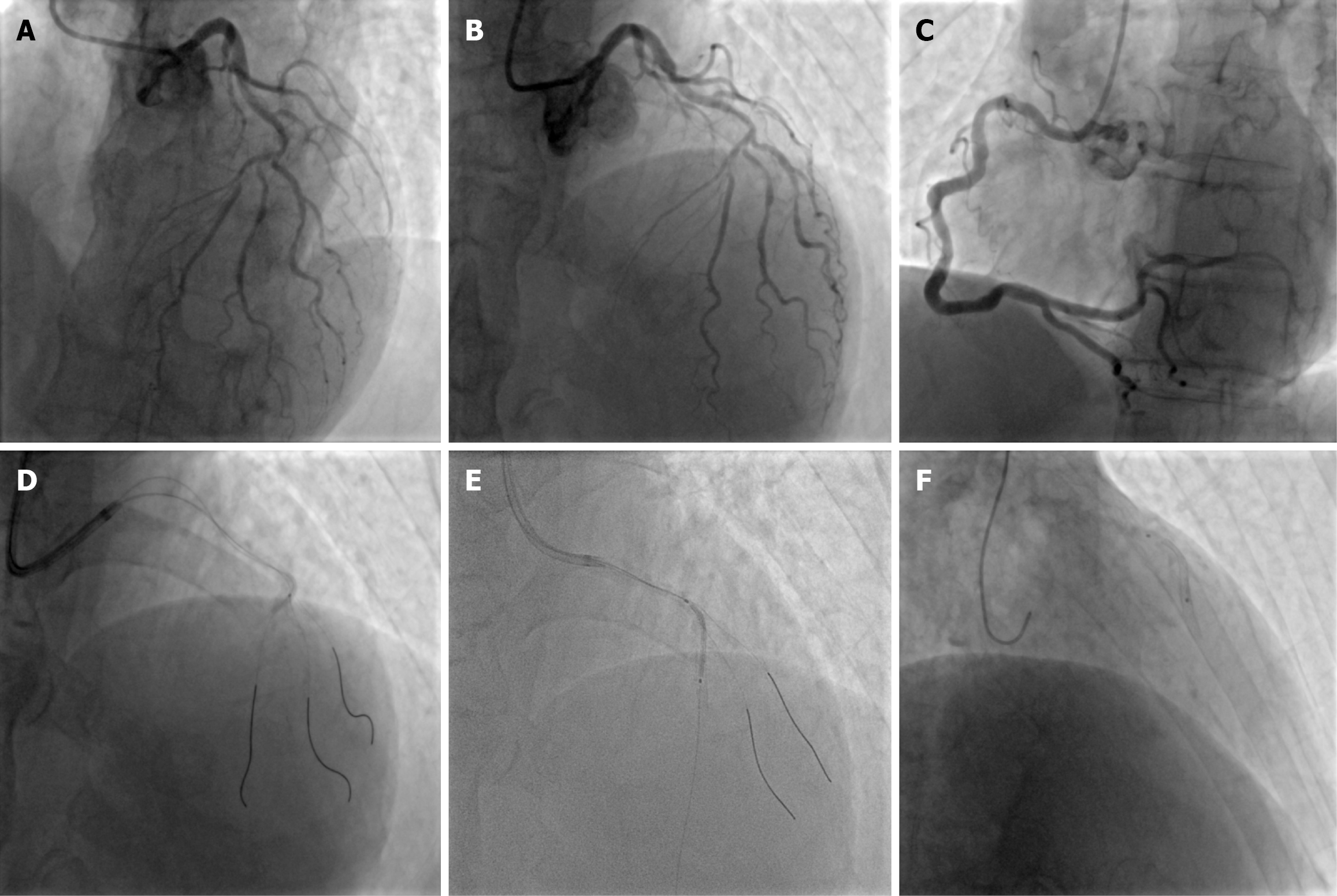
Figure 1 Coronary stent balloon shaft fracture during initial percutaneous coronary intervention.
A and B: Baseline angiography: Calcified left anterior descending artery (LAD) with D2/D3 involvement; C: Right coronary artery: No significant stenosis; D: Guidewire advancement and predilation; E: Coronary stent balloon shaft fracture extending 50 cm from mid-LAD into the ascending aorta after stent dislodgement and failed withdrawal.
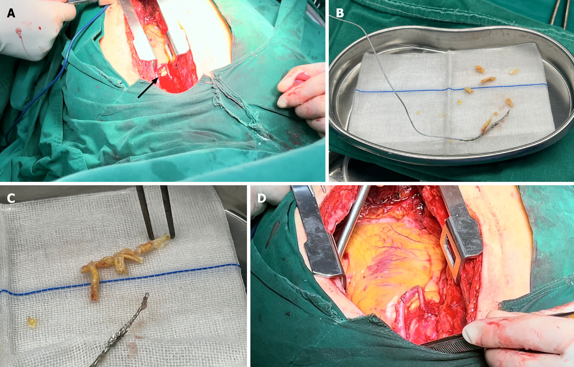
Figure 2 Surgical salvage with coronary endarterectomy.
A: Longitudinal arteriotomy of mid-left anterior descending artery (black arrowheads); B: Retrieved specimen: Coronary stent balloon fracture approximately 50 cm in length and two bare metal stents embedded in calcified tissue; C: Reconstructed plaque with protruding stent ends; D: Patch closure of arteriotomy.
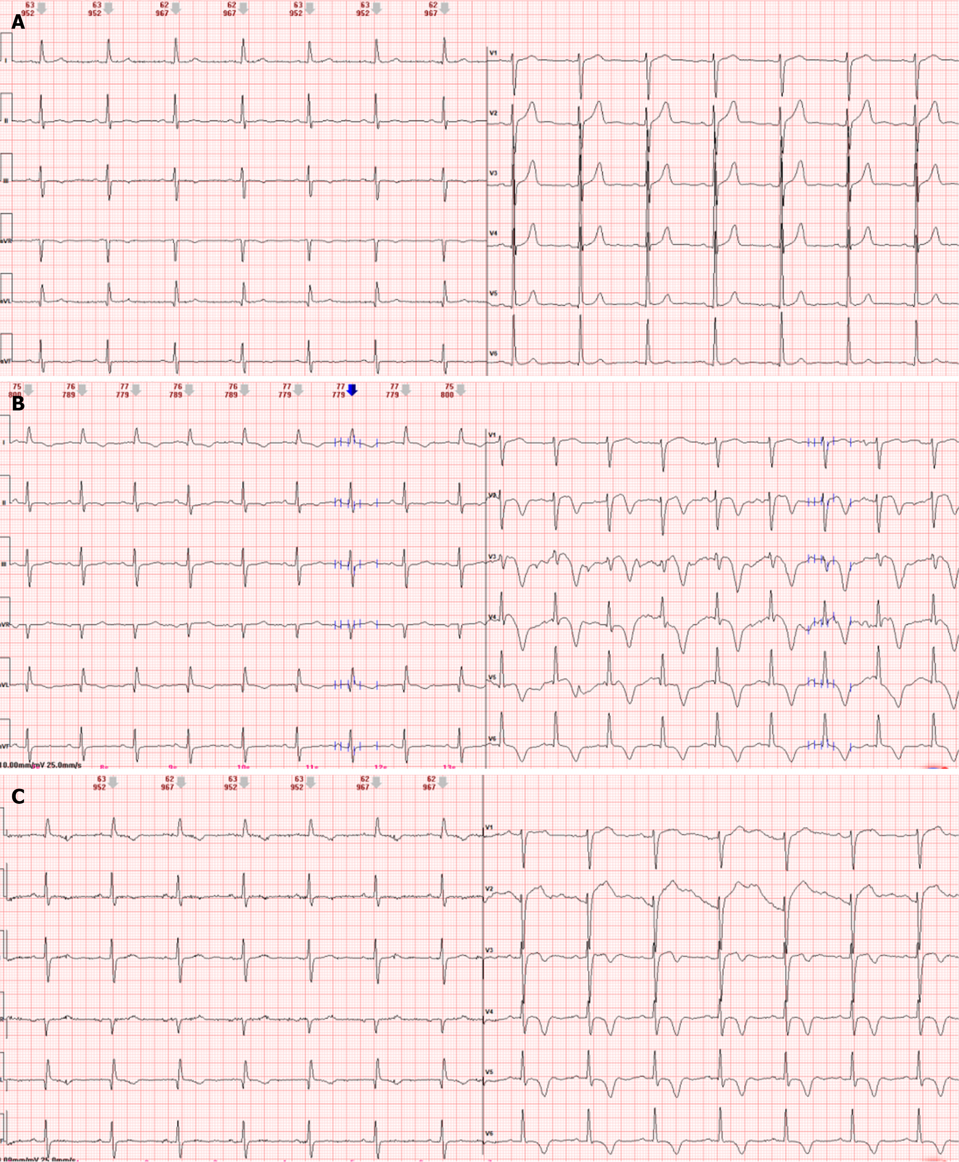
Figure 3 Serial electrocardiogram changes.
A: Admission electrocardiogram (ECG): Pseudonormalization of T waves in V2-V4; B: ECG during recurrent angina: New ST depression and deep T-wave inversions in V2-V6, confirming active ischemia; C: Post-procedural 12-lead electrocardiogram of second percutaneous coronary intervention showing significant recovery of T-waves in leads V2-V6, suggesting improved myocardial reperfusion.
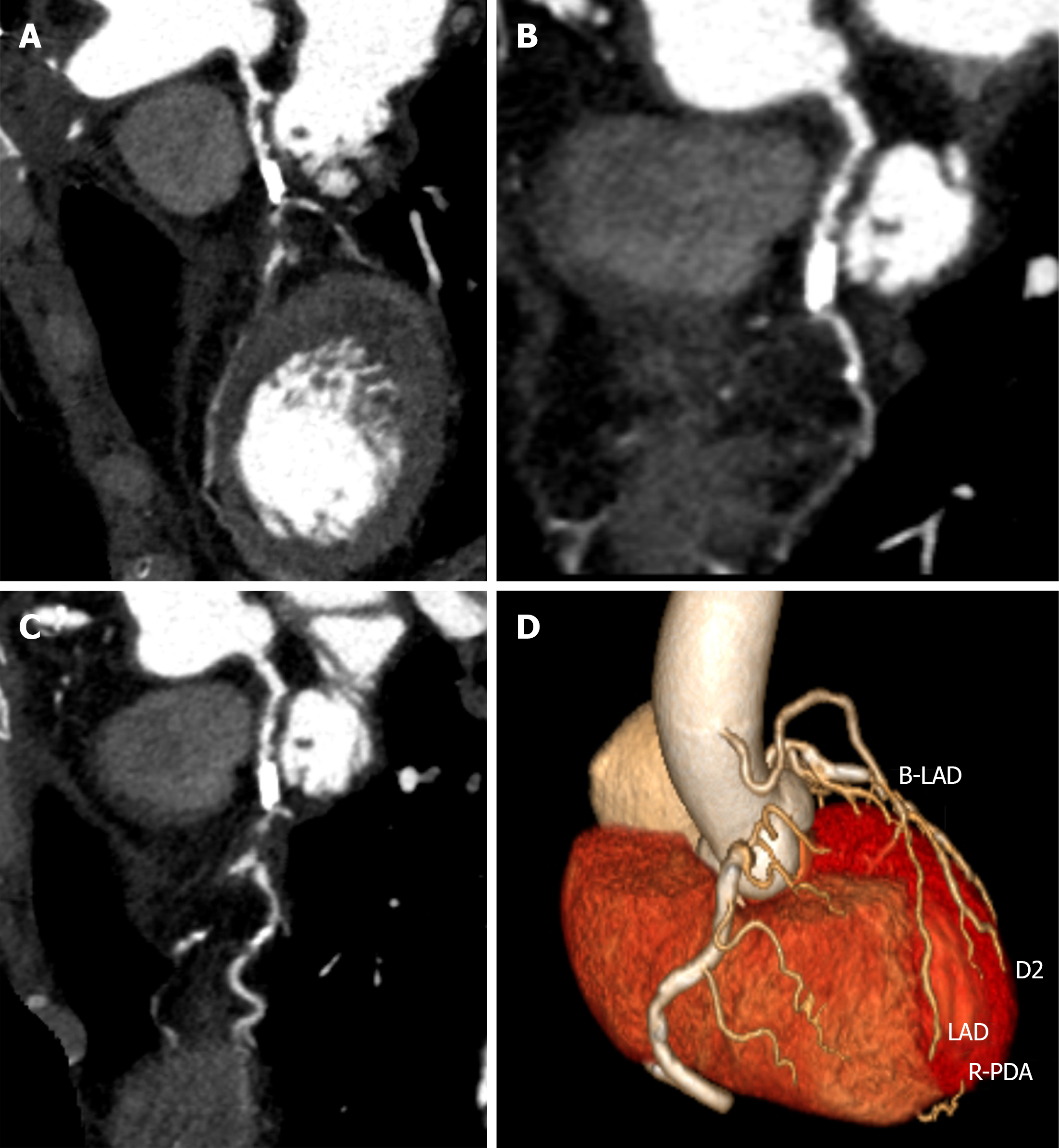
Figure 4 Coronary computed tomography angiography following coronary artery bypass grafting.
A: Left anterior descending artery (LAD): Proximal calcified plaque with diffuse narrowing, suspected mid-to-distal occlusion; B: D2: Ostial severe stenosis with mid-segment calcification; C: D3: Proximal severe stenosis with tortuosity; D: 3D reconstruction: Patent saphenous vein graft to LAD, occluded native LAD at anastomosis. B-LAD: Saphenous vein bypass graft to the left anterior descending coronary artery; D2: Second diagonal branch; D3: Third diagonal branch; LAD: Left anterior descending artery; R-PDA: Right posterior descending artery.
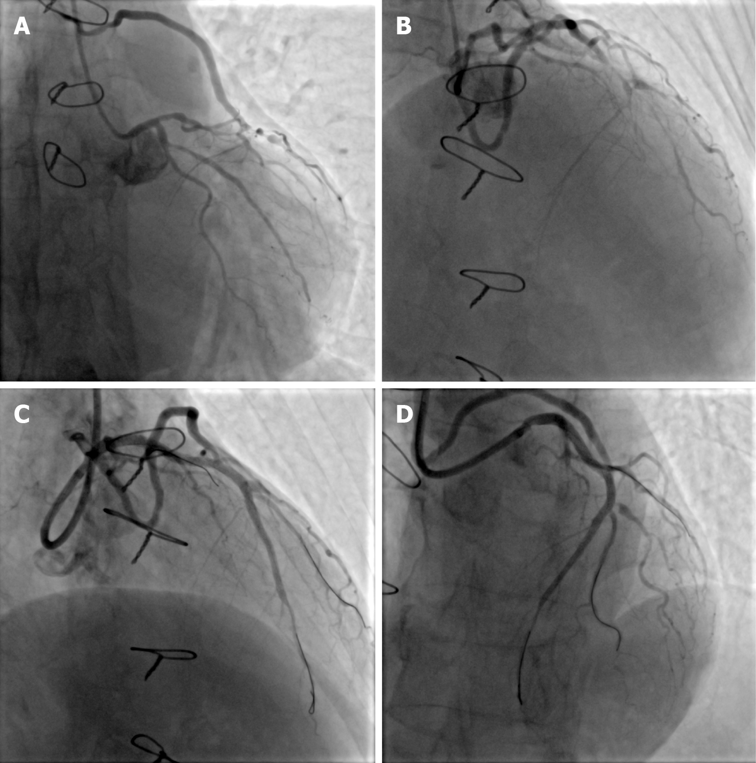
Figure 5 Re-percutaneous coronary intervention with intravascular lithotripsy rupture and successful stenting.
A and B: Baseline angiography: Patent saphenous vein graft, severe anastomotic restenosis, occluded mid-left anterior descending artery; C: Intravascular lithotripsy balloon rupture on fluoroscopy; D: Final angiography: TIMI 3 flow after three stents.
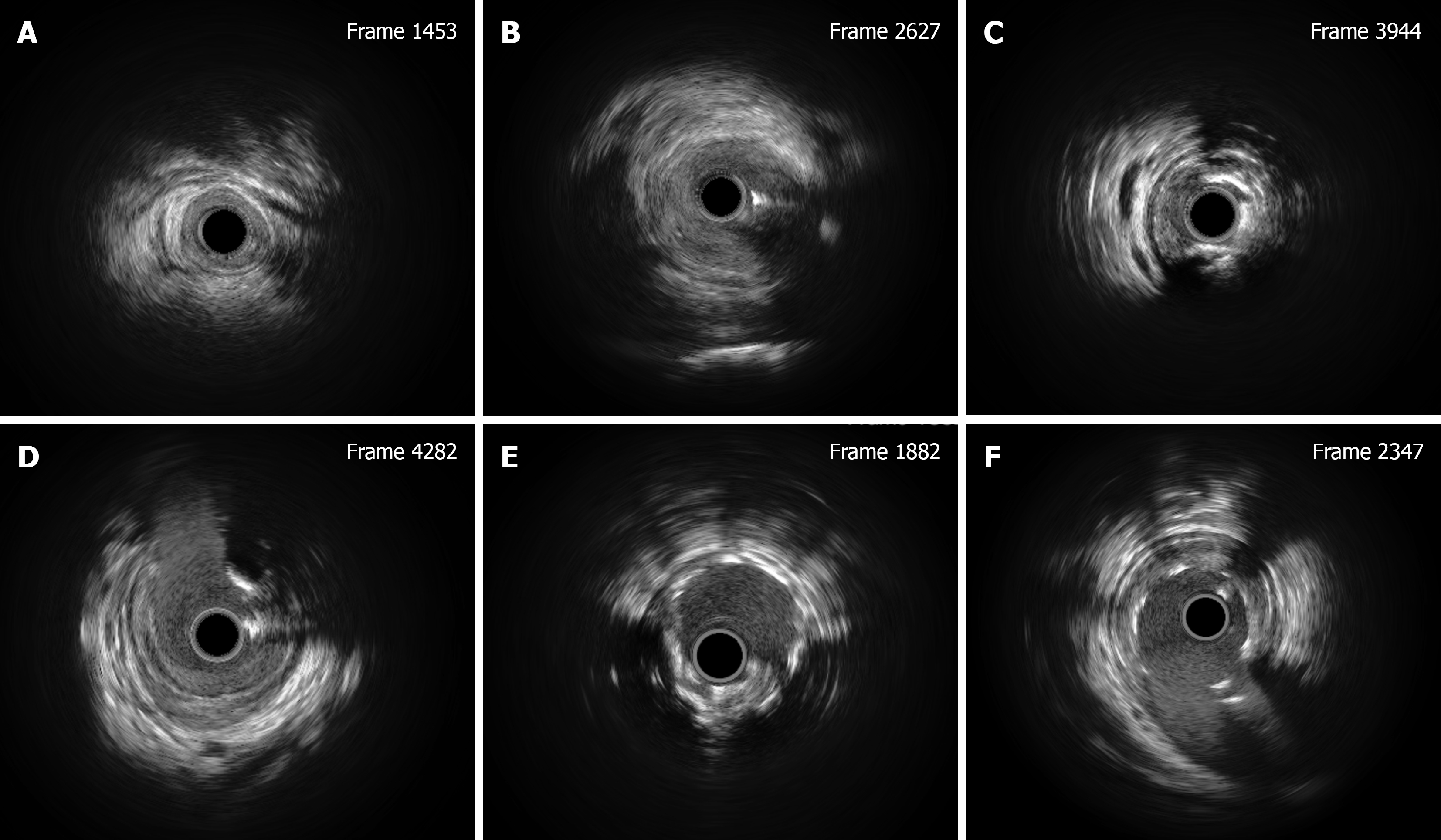
Figure 6 Intravascular ultrasound findings during re-percutaneous coronary intervention.
A: Mid-left anterior descending artery (LAD): Myocardial bridging, no plaque; B: Mid-LAD (post-endarterectomy): Hypoechoic plaques, no calcification; C: Proximal LAD: 270° circumferential calcification; D: LAD ostium: Calcified nodule (acoustic shadowing), prompting IVL; E: Full drugeluting stent implantation apposition of 270° circumferential calcification; F: Adequate drugeluting stent implantation apposition of calcified nodule.
- Citation: Huang D, Li RZ, Chen HM, He B, Yan W, Li KL, Long M. Stent balloon fracture, endarterectomy, and lithotripsy balloon rupture following percutaneous coronary intervention: A case report. World J Cardiol 2026; 18(8): 124678
- URL: https://www.wjgnet.com/1949-8462/full/v18/i8/124678.htm
- DOI: https://dx.doi.org/10.4330/wjc.124678