
















Copyright: ©Author(s) 2026.
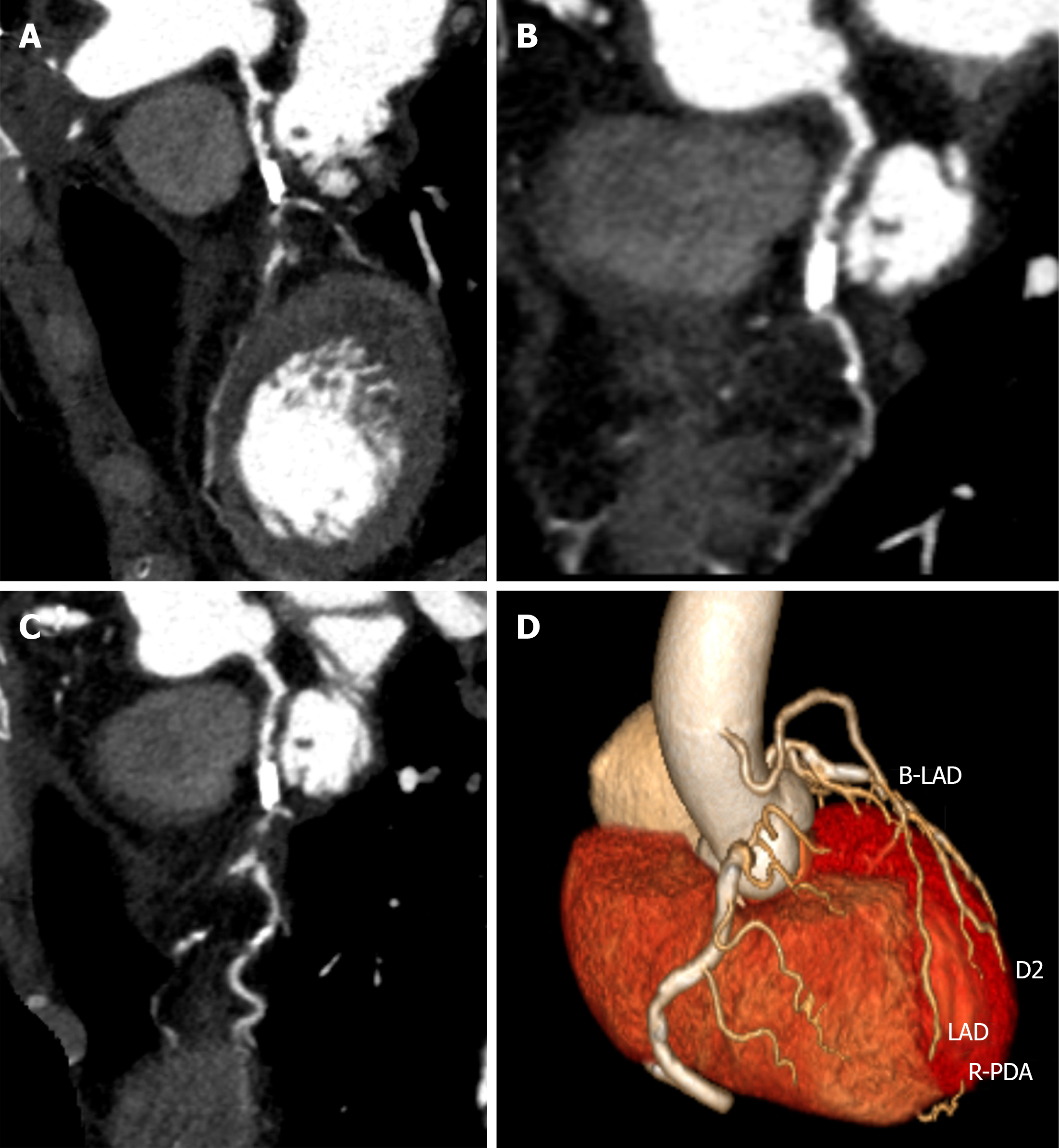
Figure 4 Coronary computed tomography angiography following coronary artery bypass grafting.
A: Left anterior descending artery (LAD): Proximal calcified plaque with diffuse narrowing, suspected mid-to-distal occlusion; B: D2: Ostial severe stenosis with mid-segment calcification; C: D3: Proximal severe stenosis with tortuosity; D: 3D reconstruction: Patent saphenous vein graft to LAD, occluded native LAD at anastomosis. B-LAD: Saphenous vein bypass graft to the left anterior descending coronary artery; D2: Second diagonal branch; D3: Third diagonal branch; LAD: Left anterior descending artery; R-PDA: Right posterior descending artery.
- Citation: Huang D, Li RZ, Chen HM, He B, Yan W, Li KL, Long M. Stent balloon fracture, endarterectomy, and lithotripsy balloon rupture following percutaneous coronary intervention: A case report. World J Cardiol 2026; 18(8): 124678
- URL: https://www.wjgnet.com/1949-8462/full/v18/i8/124678.htm
- DOI: https://dx.doi.org/10.4330/wjc.124678