Copyright
©The Author(s) 2025.
World J Gastrointest Surg. Aug 27, 2025; 17(8): 109069
Published online Aug 27, 2025. doi: 10.4240/wjgs.v17.i8.109069
Published online Aug 27, 2025. doi: 10.4240/wjgs.v17.i8.109069
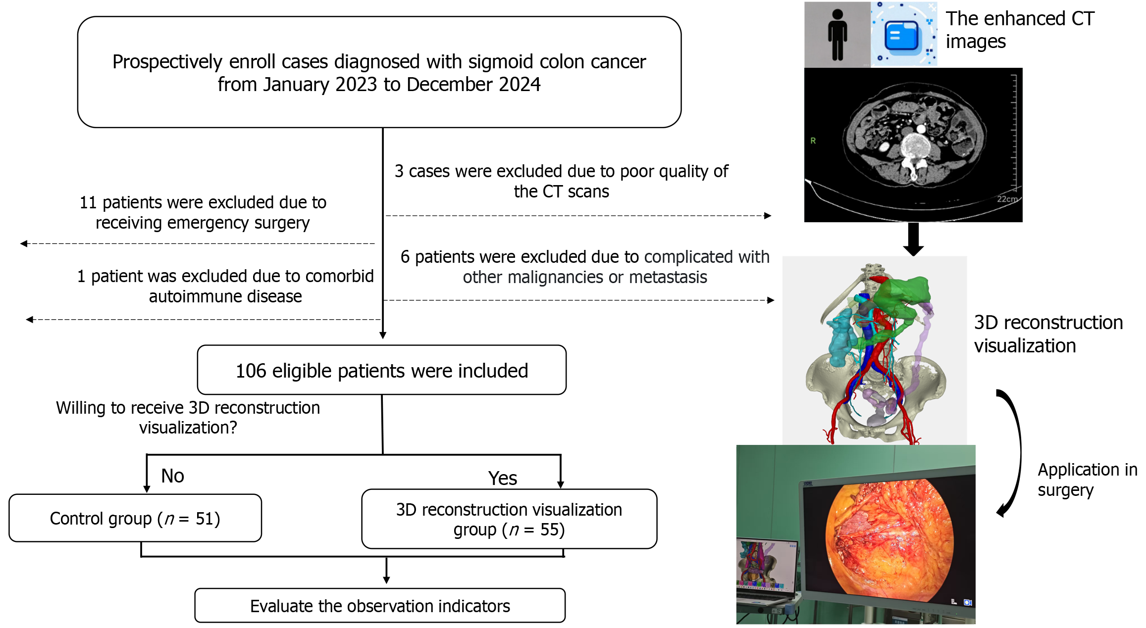
Figure 1 The design and flowchart of this study.
3D: Three-dimensional; CT: Computed tomography.
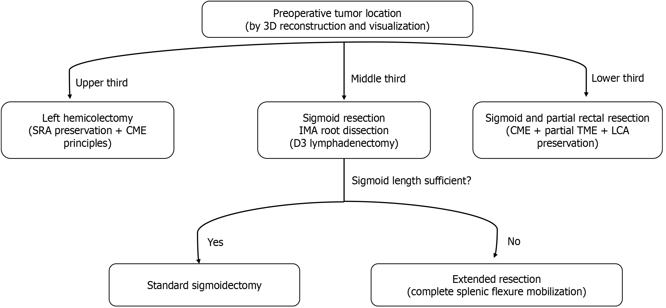
Figure 2 With the assistance of three-dimensional reconstruction and visualization, the surgical plan was determined pre-operatively based on the tumor location and the length of the sigmoid colon.
3D: Three-dimensional; SRA: Superior rectal artery; CME: Complete mesocolic excision; LCA: Left colic artery; IMA: Inferior mesenteric artery; TME: Total mesorectal excision.
Figure 3 The three-dimensional reconstruction visualization technology was applied in surgery.
A: Tumor localization and optimization of trocar placement; B: Measuring the virtual sigmoid colon length and evaluating whether mobilization of the splenic flexure of the colon is necessary (the black solid line in the figure represents the length of the sigmoid colon and the resection range, and the black dotted line represents the intestinal tract after the planned anastomosis); C: Marking the location of the lymph nodes that may have metastasized; D: Showing the classification of the inferior mesenteric artery, which is helpful for the preservation of the left colic artery or the superior rectal artery; E: Showing the key anatomical structure (the ureter) to guide the surgeon into the Toldt's space; F: Showing the marginal artery to prevent accidental intraoperative injury (as indicated by the black arrow in the figure).
- Citation: Zhao ZX, Yao RD, Hu ZJ, Chen CQ, Zhu S, Yao Y. Navigating anatomical complexity in laparoscopic sigmoid cancer surgery: A three-dimension reconstruction protocol for intraoperative safety and efficiency. World J Gastrointest Surg 2025; 17(8): 109069
- URL: https://www.wjgnet.com/1948-9366/full/v17/i8/109069.htm
- DOI: https://dx.doi.org/10.4240/wjgs.v17.i8.109069


