Copyright
©The Author(s) 2025.
World J Gastrointest Surg. Jun 27, 2025; 17(6): 106264
Published online Jun 27, 2025. doi: 10.4240/wjgs.v17.i6.106264
Published online Jun 27, 2025. doi: 10.4240/wjgs.v17.i6.106264
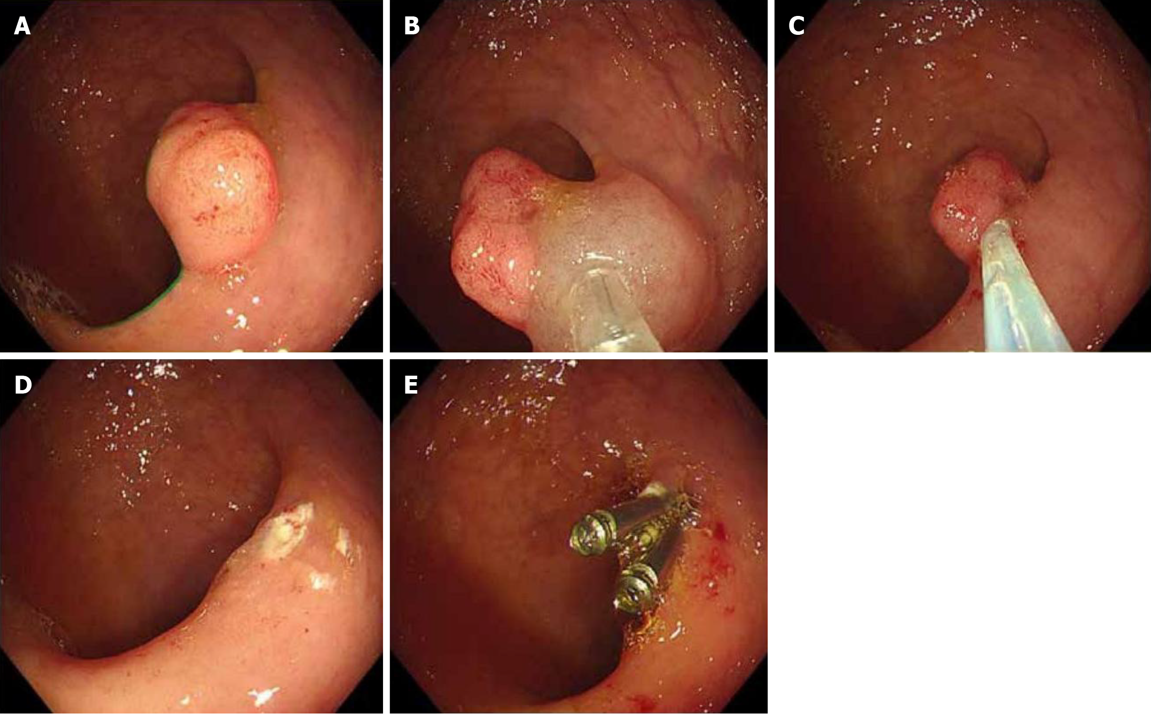
Figure 1 The procedure for colonoscopic polypectomy.
A: Electronic colonoscopy showing a colonic polyp around 1.8 cm × 1.5 cm in size located on the rectal wall; B: Multiple injections with the solution comprising glycerol fructose, epinephrine, and methylene blue to achieve complete mucosal elevation. C: Using a snare to encircle the base of the polyp, leading to its complete resection; D: Careful rechecking of the wound surface to ensure that no residual tissues remain and that the surface is smooth; E: Using three hemostatic clips to close the wound surface, preventing postoperative bleeding and infection.
- Citation: Ma YP, Zheng XY, Shen XF, Ling YT, Qian MP, Ni MJ. Impact of enhanced bowel preparation on complications and prognosis following colonoscopic polypectomy. World J Gastrointest Surg 2025; 17(6): 106264
- URL: https://www.wjgnet.com/1948-9366/full/v17/i6/106264.htm
- DOI: https://dx.doi.org/10.4240/wjgs.v17.i6.106264


