Copyright
©The Author(s) 2025.
World J Gastrointest Surg. Jun 27, 2025; 17(6): 106069
Published online Jun 27, 2025. doi: 10.4240/wjgs.v17.i6.106069
Published online Jun 27, 2025. doi: 10.4240/wjgs.v17.i6.106069
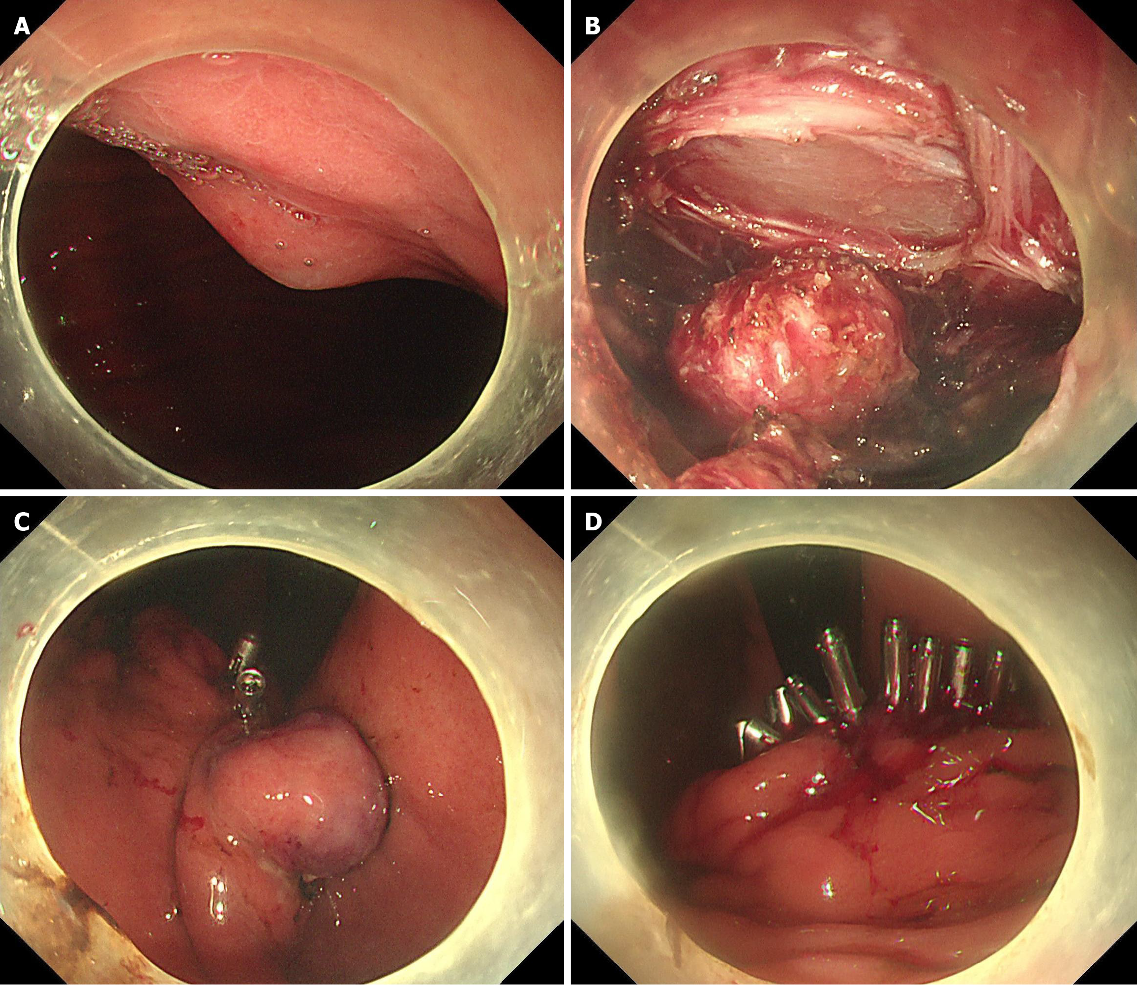
Figure 1 Resection of gastric submucosal lesions by interrupted closure in endoscopic full-thickness resection.
A: The gastric subepithelial lesion in the gastric body was initially detected by endoscopy; B: A full-thickness incision around two-thirds circumferential of the lesion; C: The metallic clips closure were performed at the proximal of the wound surface; D: The wound was completely sutured with metal clips.
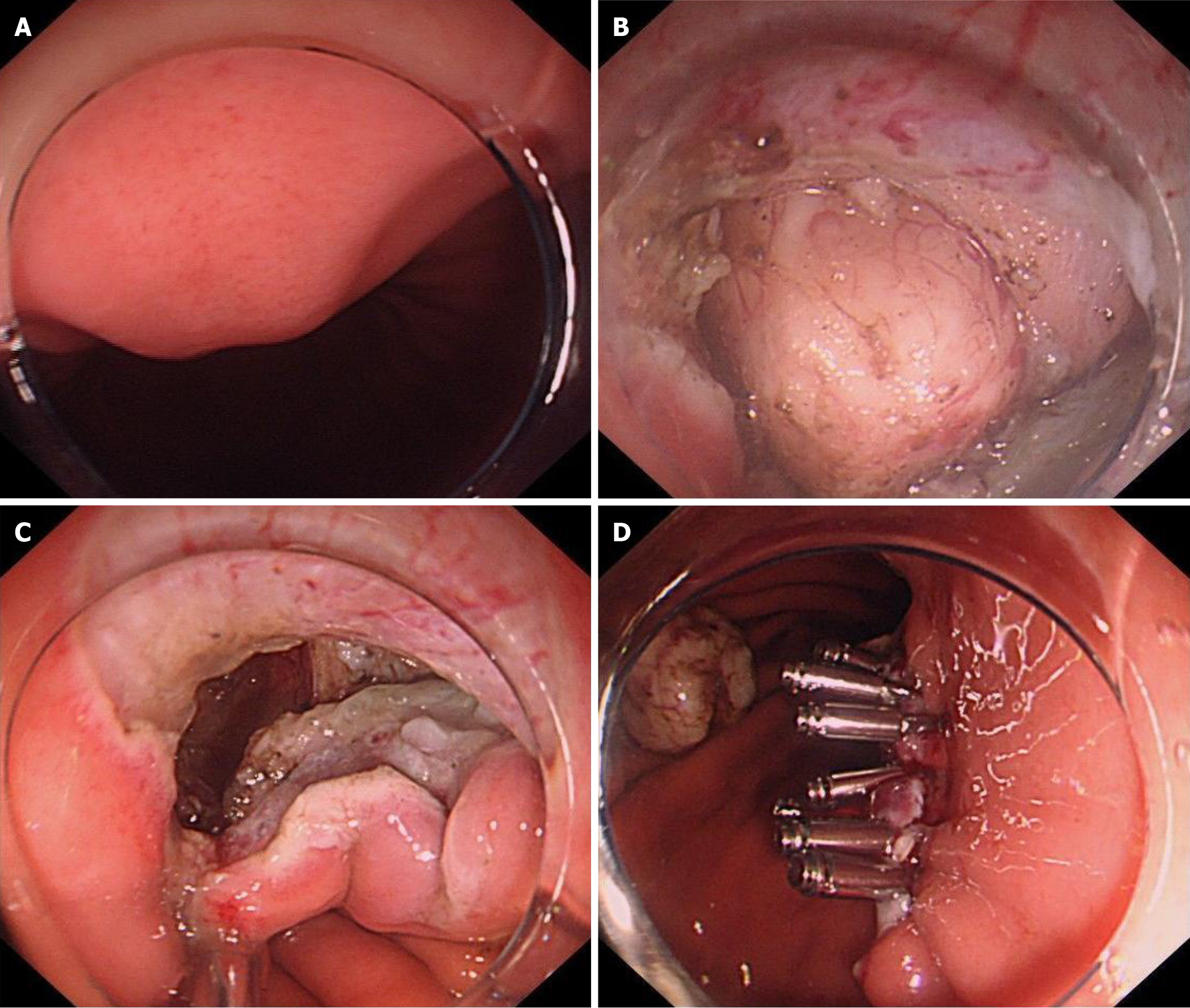
Figure 2 Resection of gastric submucosal lesions by traditional closure in endoscopic full-thickness resection.
A: The gastric subepithelial lesion in the gastric body was initially detected by endoscopy; B: The full-thickness incision around the lesion was performed; C: The gastric wall defect after the lesion resection; D: The wound was completely sutured with metallic clips.
- Citation: Zhang M, Liu J, Dong YP, Zhao Q, Lin ML, Gao TJ, Feng JL, Wang YF, Guo YF, Wang Z, Jia W, Yang Z. Comparison between interrupted closure technique and traditional closure technique in endoscopic full-thickness resection for treating gastric subepithelial lesions. World J Gastrointest Surg 2025; 17(6): 106069
- URL: https://www.wjgnet.com/1948-9366/full/v17/i6/106069.htm
- DOI: https://dx.doi.org/10.4240/wjgs.v17.i6.106069


