Copyright
©The Author(s) 2016.
World J Diabetes. Sep 15, 2016; 7(17): 406-411
Published online Sep 15, 2016. doi: 10.4239/wjd.v7.i17.406
Published online Sep 15, 2016. doi: 10.4239/wjd.v7.i17.406
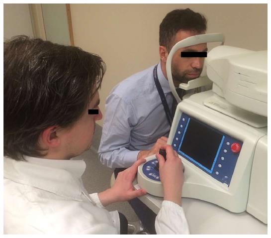
Figure 1 Acquisition of the images with in vivo corneal confocal microscopy.
Patient is comfortably seated in front of the machine whilst the operator advances the scanning probe against the cornea with a joystick.
Figure 2 Corneal innervation evaluated by in vivo corneal confocal microscopy in a health subjects (A), in a subject affected by type 1 diabetes without cardiac autonomic neuropathy (B) and in a subject affected by type 1 diabetes with cardiac autonomic neuropathy (C).
Nerve fiber density and length is reduced in people with type 1 diabetes and in those with cardiac autonomic neuropathy.
- Citation: Maddaloni E, Sabatino F. In vivo corneal confocal microscopy in diabetes: Where we are and where we can get. World J Diabetes 2016; 7(17): 406-411
- URL: https://www.wjgnet.com/1948-9358/full/v7/i17/406.htm
- DOI: https://dx.doi.org/10.4239/wjd.v7.i17.406


