Copyright
©2013 Baishideng Publishing Group Co.
World J Diabetes. Dec 15, 2013; 4(6): 290-294
Published online Dec 15, 2013. doi: 10.4239/wjd.v4.i6.290
Published online Dec 15, 2013. doi: 10.4239/wjd.v4.i6.290
Figure 1 Standard photograph.
A: 2A. Notice the intraretinal hemorrhages. If 4 quadrants have intraretinal hemorrhages of at least this magnitude then by definition severe non-proliferative retinopathy is present; B: 6A. Notice venous beading (VB). If 2 quadrants or more have VB of at least this magnitude then by definition severe non-proliferative retinopathy is present; C: 8A. Notice the intraretinal microvascular abnormalities (IRMA). If one or more quadrants has IRMA of at least this magnitude then by definition severe non-proliferative retinopathy is present.
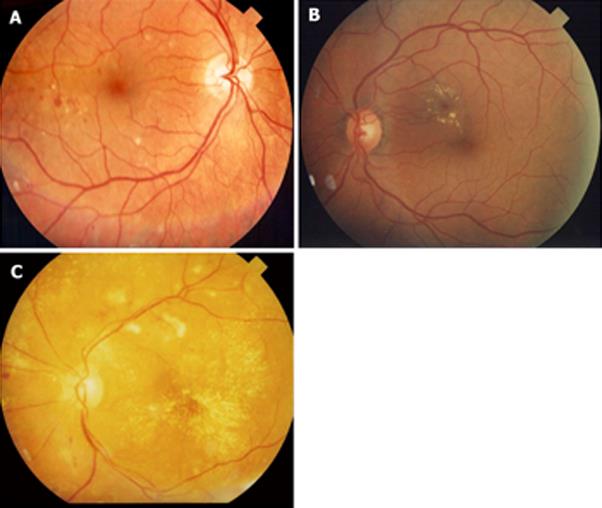
Figure 2 Diabetic macular edema.
A: Mild diabetic macular edema (DME). Notice that the hard exudates are located far from the center of the fovea; B: Moderate DME. Even though there is no thickening involving the center of the fovea, the hard exudates are threatening the center of the fovea; C: Severe DME. The center of the fovea is involved with hard exudate and thickening.
- Citation: Wu L, Fernandez-Loaiza P, Sauma J, Hernandez-Bogantes E, Masis M. Classification of diabetic retinopathy and diabetic macular edema. World J Diabetes 2013; 4(6): 290-294
- URL: https://www.wjgnet.com/1948-9358/full/v4/i6/290.htm
- DOI: https://dx.doi.org/10.4239/wjd.v4.i6.290


