Copyright
©2013 Baishideng Publishing Group Co.
World J Diabetes. Aug 15, 2013; 4(4): 101-113
Published online Aug 15, 2013. doi: 10.4239/wjd.v4.i4.101
Published online Aug 15, 2013. doi: 10.4239/wjd.v4.i4.101
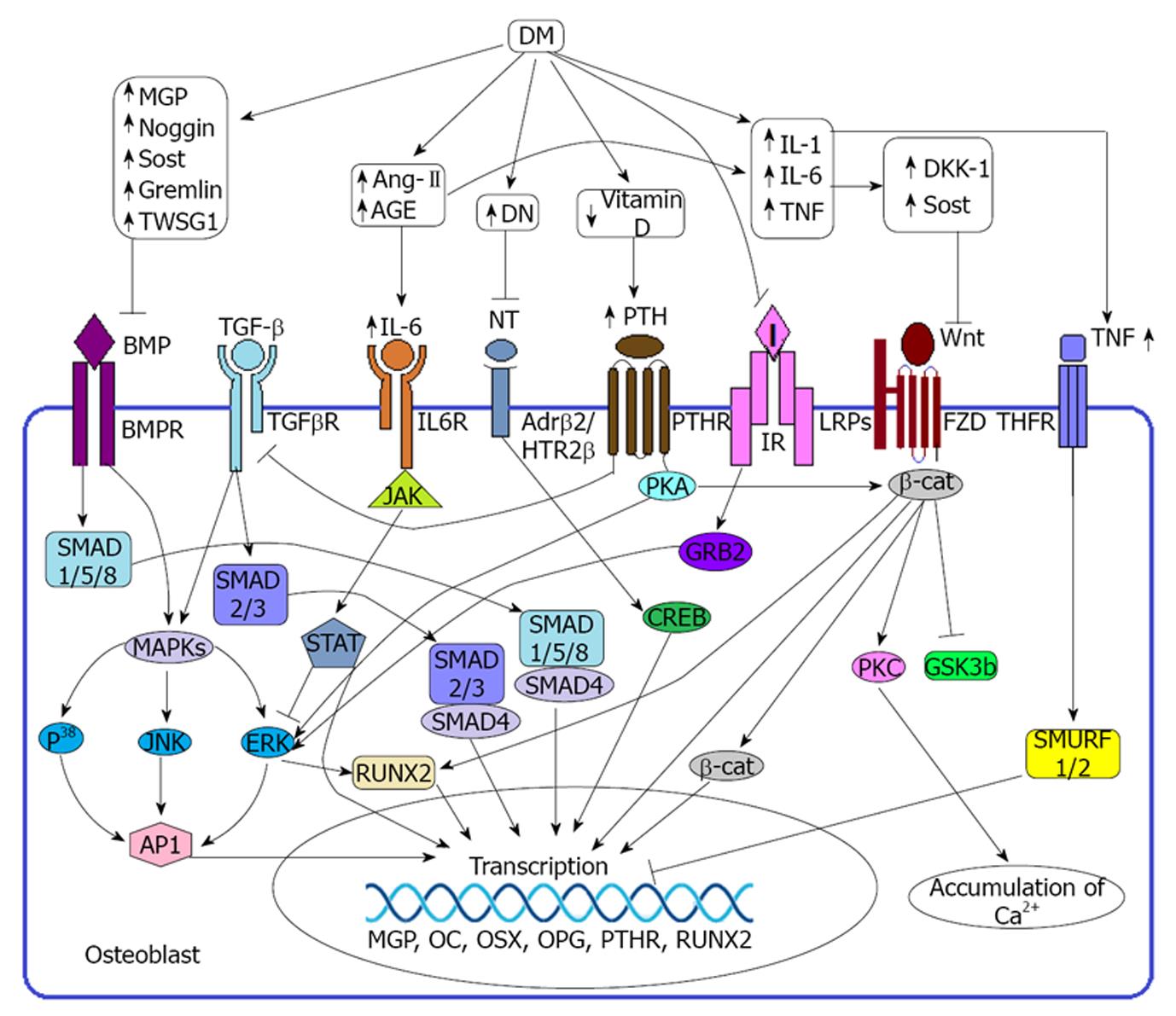
Figure 1 Diabetes mellitus induced regulation of osteoblast.
Duringhealthy condition bone morphogenetic protein (BMP), transforming growth factor β (TGF-β), Wnt, insulin and neurotransmitter signaling are mandatory to the osteoblast for its normal functioning and survival. Binding of BMP with its receptor (BMPR) activates the corresponding gene through (a) Smad depended pathway: which requires the SMADs protein (SMAD 1/5/8) or (b) non smad depended pathway: in which activates RUNX2 or AP-1 through MAPK-ERK mediated pathway. Wnt-Frizzled pathway positively regulates gene expression through β-catenin or RUNX2 mediated pathway and Calcium accumulation through PKC mediated pathway. TGF-β is also a positive regulator of osteoblast function and exerts its effect on the respective gene through SMAD 2/3 depended pathway or MAPK-ERK mediated pathway. Peripheral nerve exposure to the osteoblast signals through the adrenergic receptor 2 β (Adrβ2) or 5HTR induced pathway. Binding of neurotransmitters on the Adrβ2 or 5HTR receptor activates ERK or CREB to induce the expression of osteoblastic gene. Insulin is a beneficial factor of bone formation and it exerts its effect through GRB2-ERK mediated pathway. During diabetes mellitus (DM), hyperglycemia may induce the expression of several BMP inhibitors including MGP, Noggin, Sost, Gremlin, TWSG1 as well as several Wnt inhibitors including DKK-1, Sost. DM also induces the production of different proinflammatory cytokines including interleukin 6 (IL-6), IL-1, AT-2 and TNF which negatively regulates osteoblast functioning. Binding of IL-6 with its receptor IL-6 receptor (IL6R) sequesters ERK pathway as well as induce the gene to transcribe several inhibitors including MGP, OPG OSX. DM induced DN limits the nerve signaling through damaging the peripheral nerves. TNF binding with TNFR induce SMURF1/2 and thereby inhibit the transcription process. DM also reduces the production of vitamin D which in turn induces the secretion of parathyroid hormone (PTH). PTH binding with PTH receptor (PTHR) inhibits TGF-β signaling through inhibiting TGFβ receptor (TGFβR) although PTHR activates β-Cat and ERK pathways. DM induced IR (type-2DM) or insulin deficiency (Type-1DM) also limits insulin mediated bone formation. TNT: Neurotransmitter; HTR2β: 5-hydroxytryptamine receptor 2 β; I: Insulin; IR: Insulin receptor; LRP: Low density lipoprotein receptor related protein; FZD: Frizzled; TNF: Tumor necrosis factor; TNFR: TNF receptor; JAK: Janus kinase; STAT: Signal transducers and activators of transcription; AP-1: Activator protein 1; ERK: Extracellular signal regulated kinase; MAPK: Mitogen activated protein kinase; RUNX2: Runt related transcription factor 2; PKA: Protein kinase A; PKA: Protein kinase C; β-cat: β catenin; GSK3b: Glycogen synthase kinase 3b; SMURF: SMAD ubiquitylation regulatory factor; MGP: Matrix gla protein; OC: Osteocalcin; OSX: Osterix; OPG: Osteoprotegerin; DKK-1: Dickkopf related protein 1; Sost: Sclerostin; TWSG1: Twisted gremlin; Ang-II: Angiotensin-II; AGE: Advance glycation end product; GRB2: Growth factor receptor bound protein.
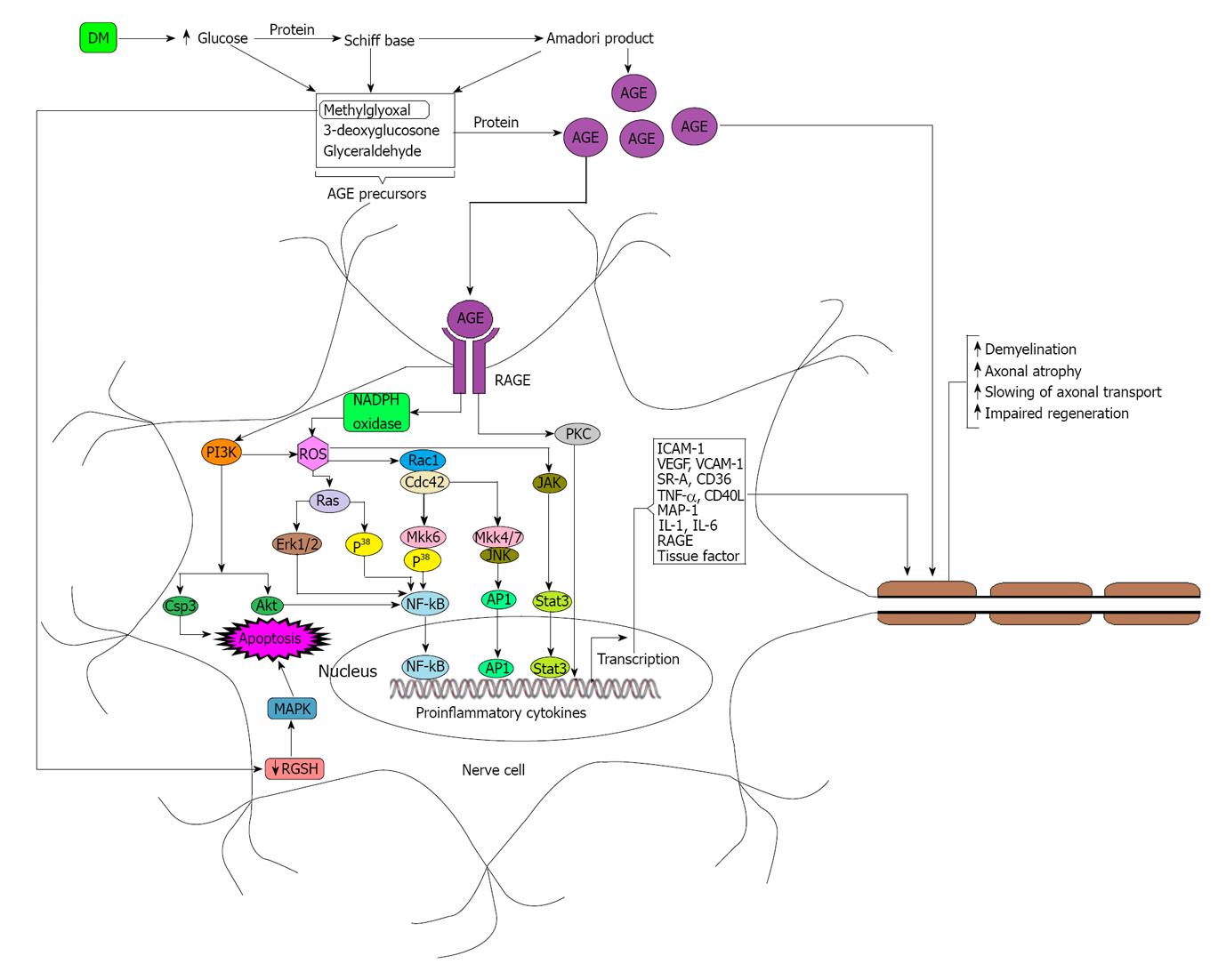
Figure 2 Diabetes mellitus induced Peripheral nerve damage.
During hyperglycemic condition concentrations of methylglyoxal, 3-deoxyglucosone and glyceraldehyde increase rapidly due to the increased breakdown of glucose. Elevated levels of methylglyoxal, 3-deoxyglucosone and glyceraldehyde lead to the formation of advance advance glycation end products (AGEs) which in turn modify nerve cell components as well as signal through the receptor for advance glycation end product (RAGE) expressed on the nerve cells in order to produce different types of cytokines which may have roles on nerve damage. RAGE induced nicotinamide adenine diphosphate hydrogen (NADPH) oxidase is the major source of reactive oxygen species (ROS) and ROS plays a crucial role to activate nuclear factor kappa B (NF-κB) through Ras-Erk, Rac1-Mkk6 depended pathway. ROS also activates AP-1 and Stat-3 through Rac1-Mkk4/7, JAK-Stat mediated pathway respectively. RAGE may induce apoptosis through PI3K-Csp3 depended pathway as well as activates NF-κB through PI3-Akt mediated pathway although PI3K may participates in ROS production. Diabetes mellitus (DM) induced methylglyoxal may directly participates in apoptosis through MAPK mediated pathway. Activated NF-κB, AP-1 and Stat3 act congruously to transcribe the genes of proinflammatory cytokines and other factors which are responsible for the destruction of peripheral nerve cells. AGE also participate directly on the modification of axon and thereby reduce the potentiality of signal transduction. DM: Diabetes mellitus; AGE: Advance glycation end product; JAK: Janus kinase; RAGE: Receptor advance glycation end product; ROS: Reactive oxygen species.
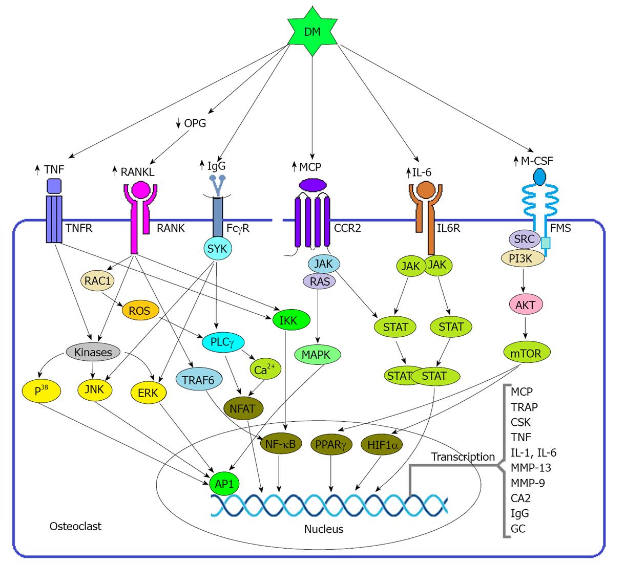
Figure 3 Diabetes mellitus induced regulation of osteoclast.
During normal physiology several osteoblastogenic modulators including RANKL, M-CSF, monocyte chemoattractant protein (MCP), and immunoglobulin G (IgG) binds with their receptors expressed on osteoclast and activates different signal transduction pathway to transcribe the particular gene. Binding of RANKL with RANK triggers several possible pathways to induce the corresponding element. It may induce transcription factor NF-κB through TRAF or IkB kinase (IKK) mediated pathway as well as induces nuclear factor of activated T cells (NFAT) through reactive oxygen species (ROS)- phospholipase Cγ (PLCγ) mediated pathway. RANK also may induce AP-1 through triggering the kinase enzymes. Macrophage colony stimulating factor (M-CSF) activates transcription factors peroxisome proliferator activated receptor γ (PPARγ) and hypoxia inducible factor 1 α (HIF1α) through PI3K-AKT mediated pathway. MCP activates AP-1 signaling through RAS-MAPK mediated pathway which requires the assistance of JAK. IgG also signals through the Fc receptor γ chain (FcγR) to activate NFAT via the induction of PLCγ as well as activates AP-1 through the kinase enzyme systems and both of the pathways require the activation of SYK. \During the state of DM, it induces the upregulation of osteoclastogenic factors stated above and thereby induce the differentiation and activity of osteoclast. In addition to the above factors, DM also induces the synthesis of some proinflammatory cytokines which also favor the bone resorption by osteoclast. interleukin 6 (IL-6) exerts its effect through JAK-STAT mediated pathway although MCP activated JAK may contribute to the activation of STAT to some extent. TNF also activates NF-κB and AP-1 through IKK and Kinase system respectively. CCR2: CC chemokine receptor 2; mTOR: Mammalian target of rapamycin; OPG: Osteoprotegerin; ERK: Extracellular signal regulated kinase; JNK: JUN N terminal kinase; TRAP: Tartrate resistant acid phosphatase; CSK: Cathepsin K; MMP: Matrix metalloproteinase; CA2: Carbonic anhydrase 2; GC: Glucocorticoid.
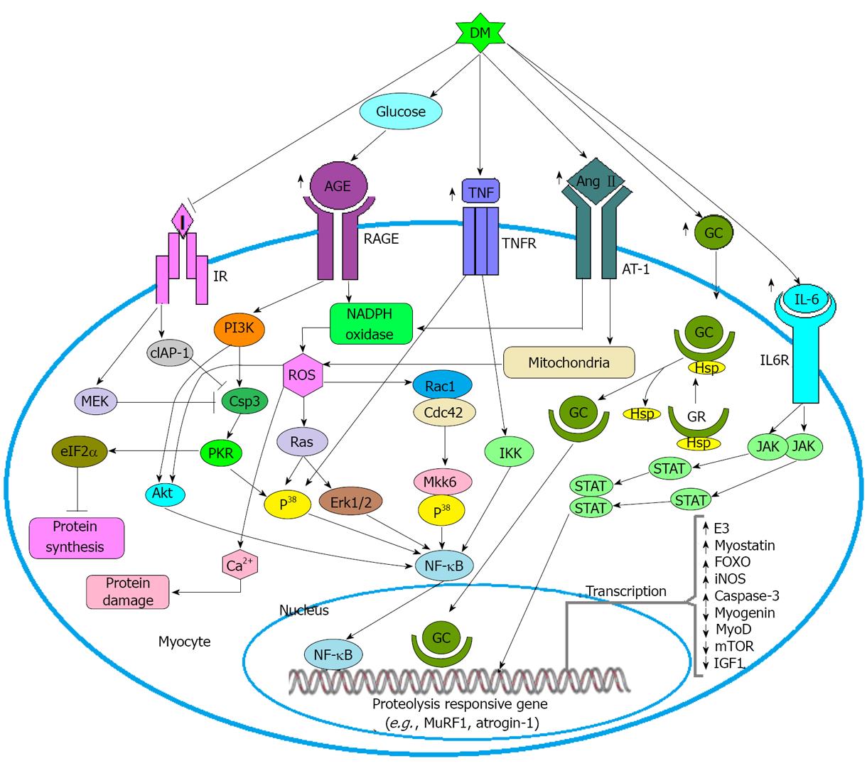
Figure 4 Diabetes mellitus induced regulation of skeletal muscle.
Diabetes mellitus (DM) induced elevated blood glucose is the major source of advance advance glycation end product (AGE) which binds with its receptor advance glycation end product (RAGE) to activate the signal cascade into myocyte. RAGE activation enhances the generation of reactive oxygen species (ROS) through the activation of nicotinamide adenine diphosphate hydrogen (NADPH) oxidase. Ang-II also induce the production ROS not only by activating NADPH oxidase but also by inducing the mitochondria. ROS may exert its effects on nuclear factor kappa B (NF-κB) through Rac1-Mkk6 and Ras mediated pathway or accelerate the damage of muscle protein through Ca2+ depended pathway. Beyond the generation of ROS, RAGE also activates PI3K which in turn activates NF-κB through Csp3- PKR and Akt mediated pathway. Activated PKR may induce the activation of eIF2α that inhibits protein synthesis. DM induced Proinflammatory cytokines interleukin 6 (IL-6) activates the gene through JAK-STAT signaling pathway and TNF activates the factor NF-κB via IKK or MAPKP38 induced pathway. Ang-II induced GC also have role in muscle atrophy and GC exerts its effect through GC-GCR complex mediated pathway. Insulin signaling is also important for muscle growth because it sequesters the activity of Csp3 through inducing the production of cIAP-1 and MEK which are potential inhibitor of Cap3. Type 1 DM reduces the production of insulin and type 2 DM makes the cell insulin resistance, so due to the deficiency of insulin it limits the functioning of cIAP-1 and MEK.
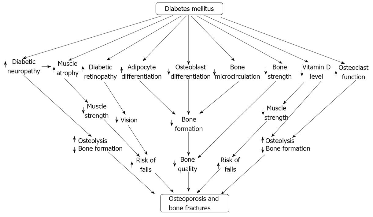
Figure 5 Possible pathways of diabetes mellitus induced osteoporosis.
- Citation: Roy B. Biomolecular basis of the role of diabetes mellitus in osteoporosis and bone fractures. World J Diabetes 2013; 4(4): 101-113
- URL: https://www.wjgnet.com/1948-9358/full/v4/i4/101.htm
- DOI: https://dx.doi.org/10.4239/wjd.v4.i4.101


