Copyright
©The Author(s) 2025.
World J Diabetes. Jul 15, 2025; 16(7): 107187
Published online Jul 15, 2025. doi: 10.4239/wjd.v16.i7.107187
Published online Jul 15, 2025. doi: 10.4239/wjd.v16.i7.107187
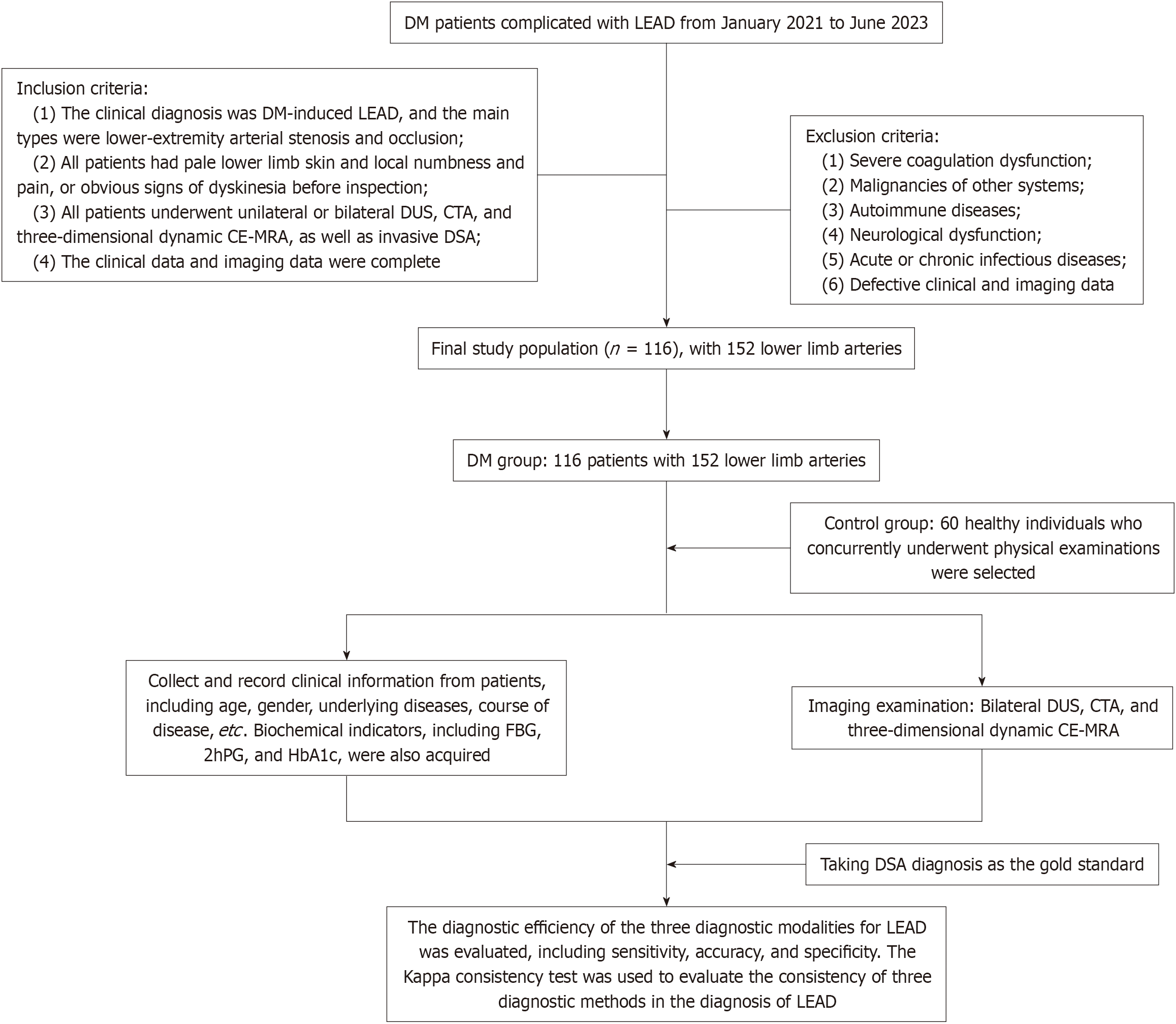
Figure 1 Flow chart of the study.
DM: Diabetes mellitus; LEAD: Lower extremity vascular diseases; DUS: Doppler ultrasonography; CTA: CT angiography; CE-MRA: Contrast-enhanced magnetic resonance angiography; DSA: Digital subtraction angiography; FBG: Fasting blood glucose; 2hPG: 2-h postprandial blood glucose; HbA1c: Glycosylated hemoglobin.
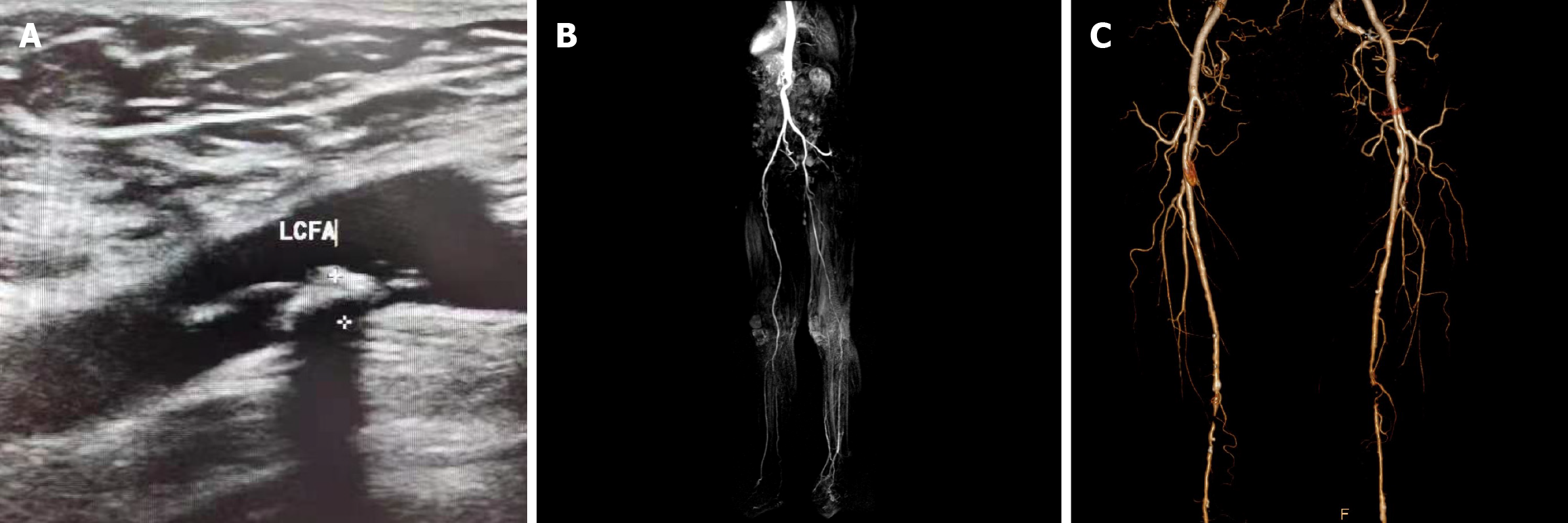
Figure 2 Patient imaging findings.
A: Doppler ultrasonography showed occlusion of the lateral femoral circumflex artery; B: Contrast-enhanced magnetic resonance angiography diagnosis results; C: Volume rendering after CT angiography examination. LCFA: Lateral circumflex femoral artery.
- Citation: Bo Y, Xie J, Xu F, Yang G, Li DL, Yan XH. Comparison of three diagnostic imaging modalities for use in diabetic inferior arterial lesions. World J Diabetes 2025; 16(7): 107187
- URL: https://www.wjgnet.com/1948-9358/full/v16/i7/107187.htm
- DOI: https://dx.doi.org/10.4239/wjd.v16.i7.107187


