Copyright
©The Author(s) 2017.
World J Gastrointest Oncol. Jul 15, 2017; 9(7): 308-313
Published online Jul 15, 2017. doi: 10.4251/wjgo.v9.i7.308
Published online Jul 15, 2017. doi: 10.4251/wjgo.v9.i7.308
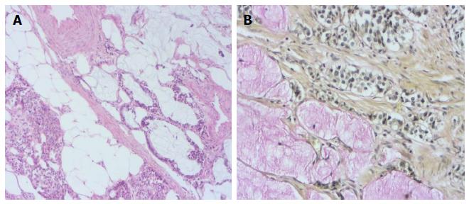
Figure 1 Heamatoxylin and eosin stained sections showing the tumor itself is composed of small uniform tumor nests of goblet cells and signet ring cells, often arranged in a microglandular fashion and sometimes accompanied by extracellular mucus (A: H and E; × 200) and adenocarcinoid tumor of goblet cell type showing positivity intra- and extra-cellular mucin deposition (B: Mucicarmine; × 400).
H and E: Heamatoxylin and eosin.
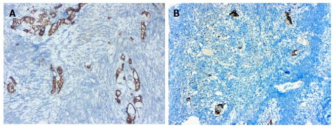
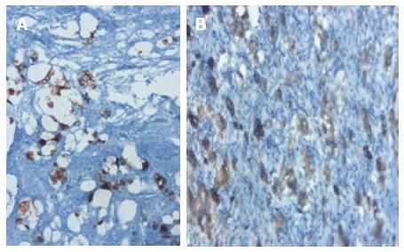
Figure 2 Tumor cells exhibit strong immunoreactivity for epithelial membrane antigen (A) and for neuroendocrine markers (B).
A: EMA, IHC; × 200; B: Chromogranin-A, IHC; × 200.
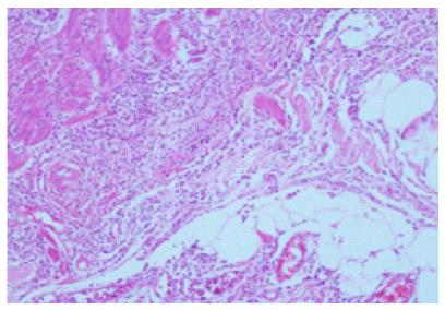
Figure 3 Adenocarcinoid tumor of goblet cell type tumor showing strong acute appendicitis findings (H and E; × 100).
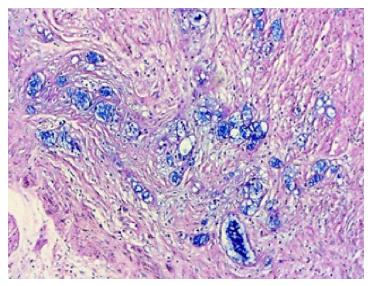
Figure 4 PAS-Alcian Blue pH 2.
5 deposition were detected in the tumor tissue (PAS/AB; × 200).
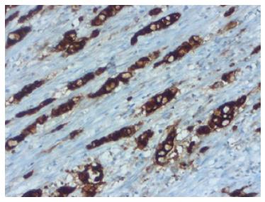
Figure 5 Adenocarcinoid tumor of goblet cell type showing positivity for immunoreactivity monoclonal carcinoembryonic antigen (IHC; × 200).
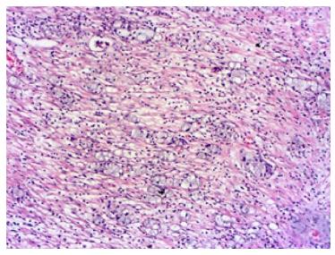
Figure 6 Heamatoxylin and eosin stained sections showing the tumor was wide, eosinophilic, had granular cytoplasm forming small nests and cords, and was characterized by small uniform nests with eccentric nuclei, signet-ring with micro glandular development and cells in goblet cell appearance (H and E; × 200).
Figure 7 In the immunohistochemical studies neuroendocrine cell component markers, synaptophysin and chromogranin-A strong focal positivity were detected in the tumor (IHC; × 200).
- Citation: Karaman H, Şenel F, Güreli M, Ekinci T, Topuz Ö. Goblet cell carcinoid of the appendix and mixed adenoneuroendocrine carcinoma: Report of three cases. World J Gastrointest Oncol 2017; 9(7): 308-313
- URL: https://www.wjgnet.com/1948-5204/full/v9/i7/308.htm
- DOI: https://dx.doi.org/10.4251/wjgo.v9.i7.308


