Copyright
©The Author(s) 2017.
World J Gastrointest Oncol. Jul 15, 2017; 9(7): 308-313
Published online Jul 15, 2017. doi: 10.4251/wjgo.v9.i7.308
Published online Jul 15, 2017. doi: 10.4251/wjgo.v9.i7.308
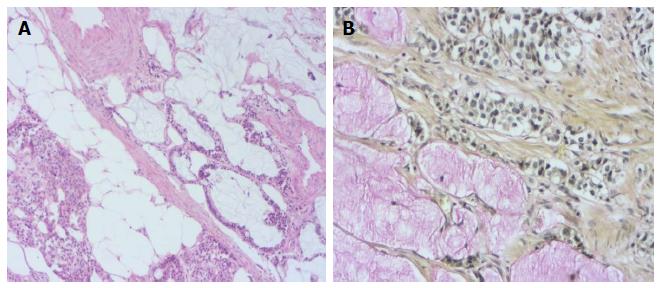
Figure 1 Heamatoxylin and eosin stained sections showing the tumor itself is composed of small uniform tumor nests of goblet cells and signet ring cells, often arranged in a microglandular fashion and sometimes accompanied by extracellular mucus (A: H and E; × 200) and adenocarcinoid tumor of goblet cell type showing positivity intra- and extra-cellular mucin deposition (B: Mucicarmine; × 400).
H and E: Heamatoxylin and eosin.
- Citation: Karaman H, Şenel F, Güreli M, Ekinci T, Topuz Ö. Goblet cell carcinoid of the appendix and mixed adenoneuroendocrine carcinoma: Report of three cases. World J Gastrointest Oncol 2017; 9(7): 308-313
- URL: https://www.wjgnet.com/1948-5204/full/v9/i7/308.htm
- DOI: https://dx.doi.org/10.4251/wjgo.v9.i7.308


