Copyright
©The Author(s) 2015.
World J Gastrointest Oncol. Aug 15, 2015; 7(8): 123-127
Published online Aug 15, 2015. doi: 10.4251/wjgo.v7.i8.123
Published online Aug 15, 2015. doi: 10.4251/wjgo.v7.i8.123
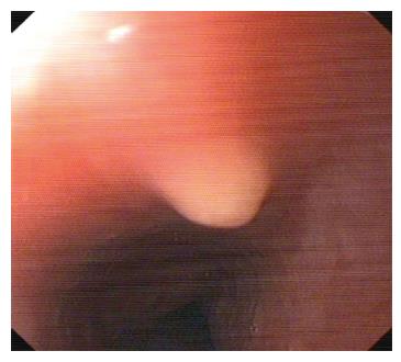
Figure 1 A grayish-yellow uplift was seen in the esophagus 18 cm from the incisor with smooth surface under endoscope in Case 4.
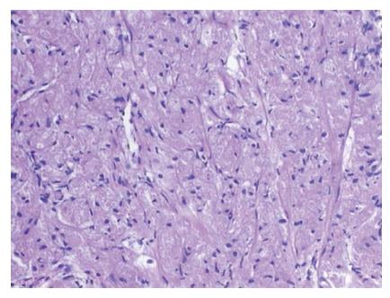
Figure 2 The tumor cells were large and polygonal with granular and eosinophilic cytoplasm, and small oval nuclei.
Hematoxylin and eosin staining, × 100.
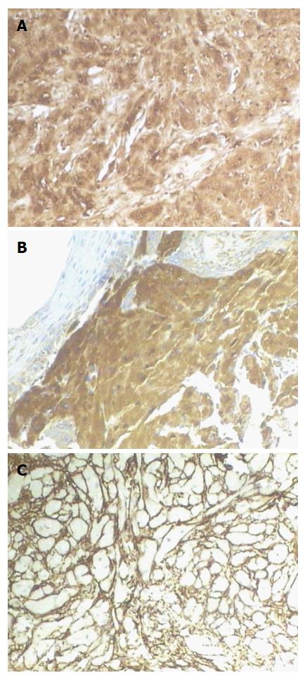
Figure 3 Immunohistochemical and histochemical staining.
Envision method, × 100. A: S-100 was strongly positive in tumor cells; B: CD68 was strongly positive in tumor cells; C: CD34 was negative in tumor cells, but the surrounding mesenchymal cells were positive for CD34.
- Citation: Wang HQ, Liu AJ. Esophageal granular cell tumors: Case report and literature review. World J Gastrointest Oncol 2015; 7(8): 123-127
- URL: https://www.wjgnet.com/1948-5204/full/v7/i8/123.htm
- DOI: https://dx.doi.org/10.4251/wjgo.v7.i8.123


