
















Copyright: ©Author(s) 2026.
World J Gastrointest Oncol. Oct 15, 2026; 18(10): 123261
Published online Oct 15, 2026. doi: 10.4251/wjgo.123261
Published online Oct 15, 2026. doi: 10.4251/wjgo.123261
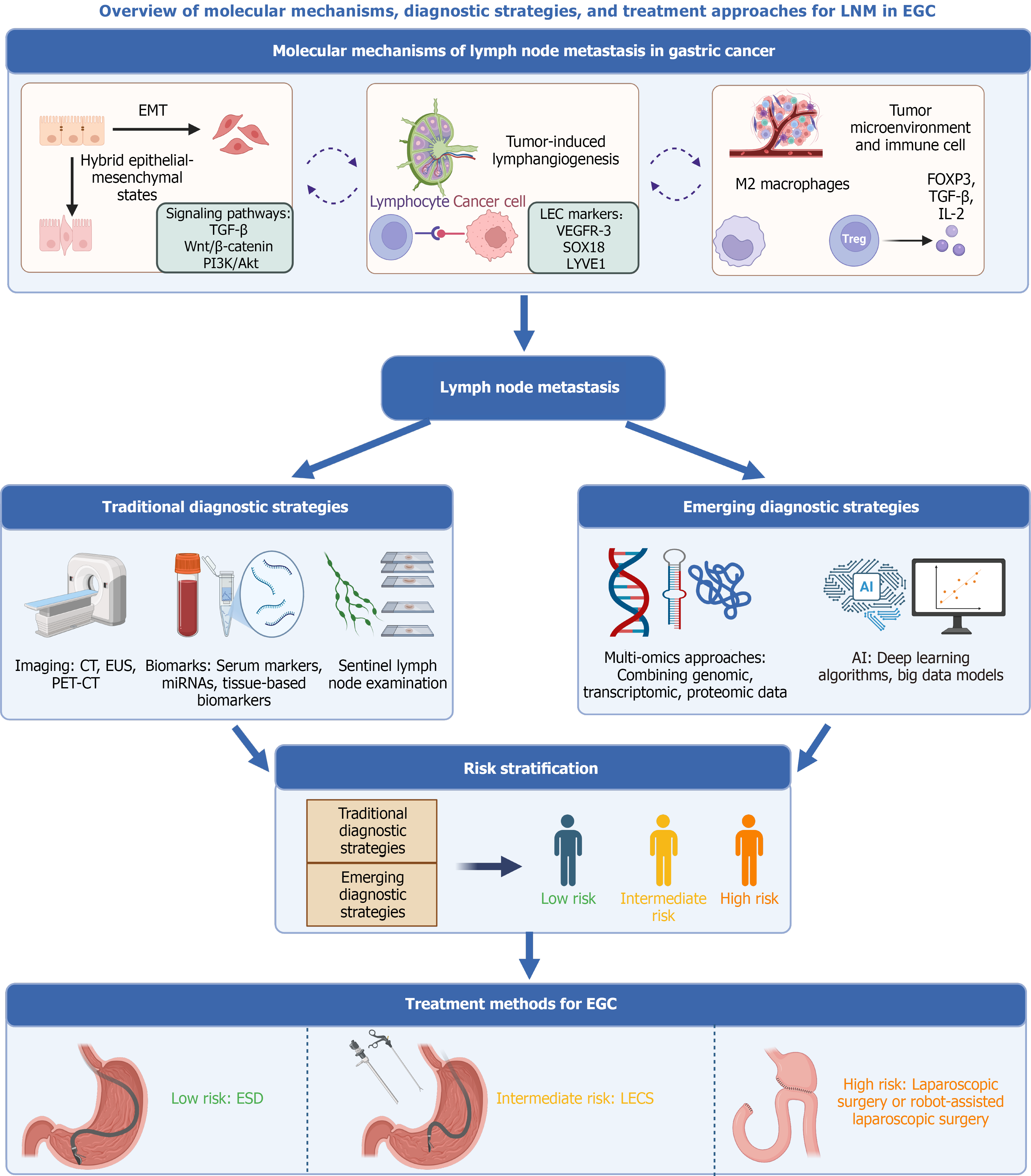
Figure 1 Overview of lymph node metastasis in early gastric cancer.
Lymph node metastasis is driven by the interaction of epithelial-mesenchymal transition, tumor-induced lymphangiogenesis, and immune regulation within the tumor microenvironment. Preoperative evaluation integrates imaging, sentinel lymph node assessment, biomarkers, and artificial intelligence models. This integrated assessment supports risk stratification and guides treatment selection, including endoscopic submucosal dissection, surgery, and laparoscopic and endoscopic cooperative surgery. LNM: Lymph node metastasis; EGC: Early gastric cancer; EMT: Epithelial-mesenchymal transition; TGF: Transforming growth factor; PI3K/Akt: Phosphatidylinositol 3’-kinase/protein kinase B; LEC: Lymphatic endothelial cells; VEGFR: Vascular endothelial growth factor receptor; IL: Interleukin; FOXP3: Forkhead box P3; Treg: Regulatory T cells; CT: Computed tomography; EUS: Endoscopic ultrasound; PET-CT: Positron emission tomography-computed tomography; miRNA: MicroRNA; AI: Artificial intelligence; ESD: Endoscopic submucosal dissection; LECS: Laparoscopic and endoscopic cooperative surgery.
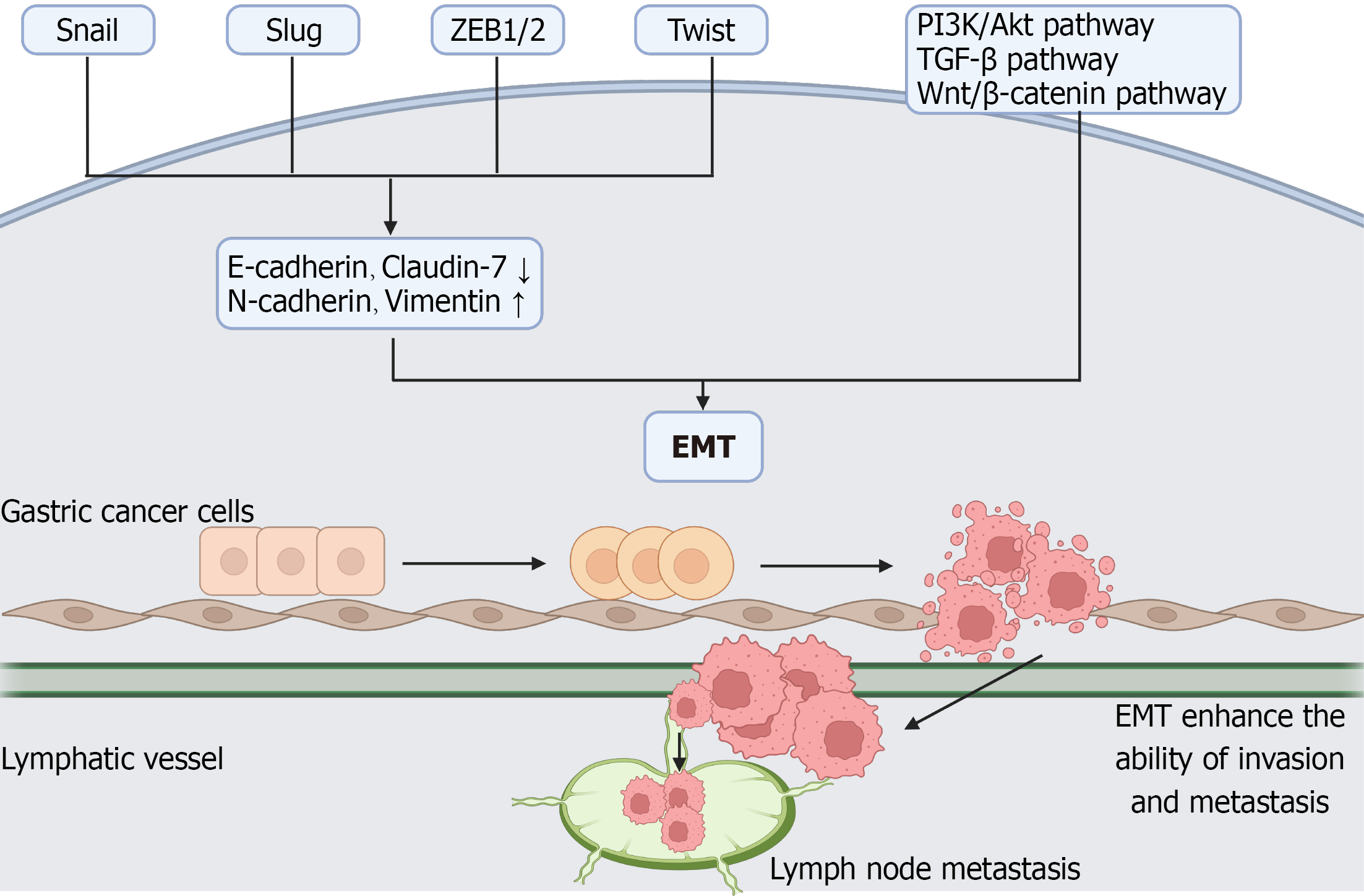
Figure 2 Schematic representation of epithelial-mesenchymal transition-driven lymph node metastasis in early gastric cancer.
Epithelial-mesenchymal transition is characterized by loss of epithelial markers and gain of mesenchymal markers, accompanied by activation of transcription factors such as Snail, Slug, zinc finger E-box binding homeobox 1/2, and Twist. These changes enhance tumor cell motility and invasiveness, facilitating tumor cell dissemination and lymphatic spread. ZEB1/2: Zinc finger E-box binding homeobox 1/2; PI3K/Akt: Phosphatidylinositol 3’-kinase/protein kinase B; TGF: Transforming growth factor; EMT: Epithelial-mesenchymal transition.
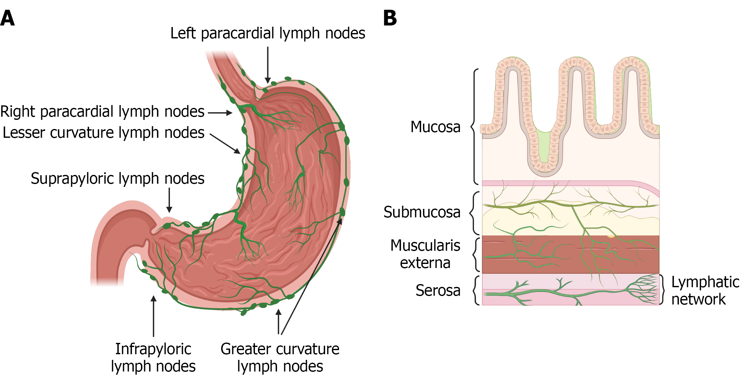
Figure 3 Anatomical basis of lymphatic dissemination in early gastric cancer.
A: Distribution of perigastric lymph nodes; B: Distribution of lymphatic vessels in the gastric wall.
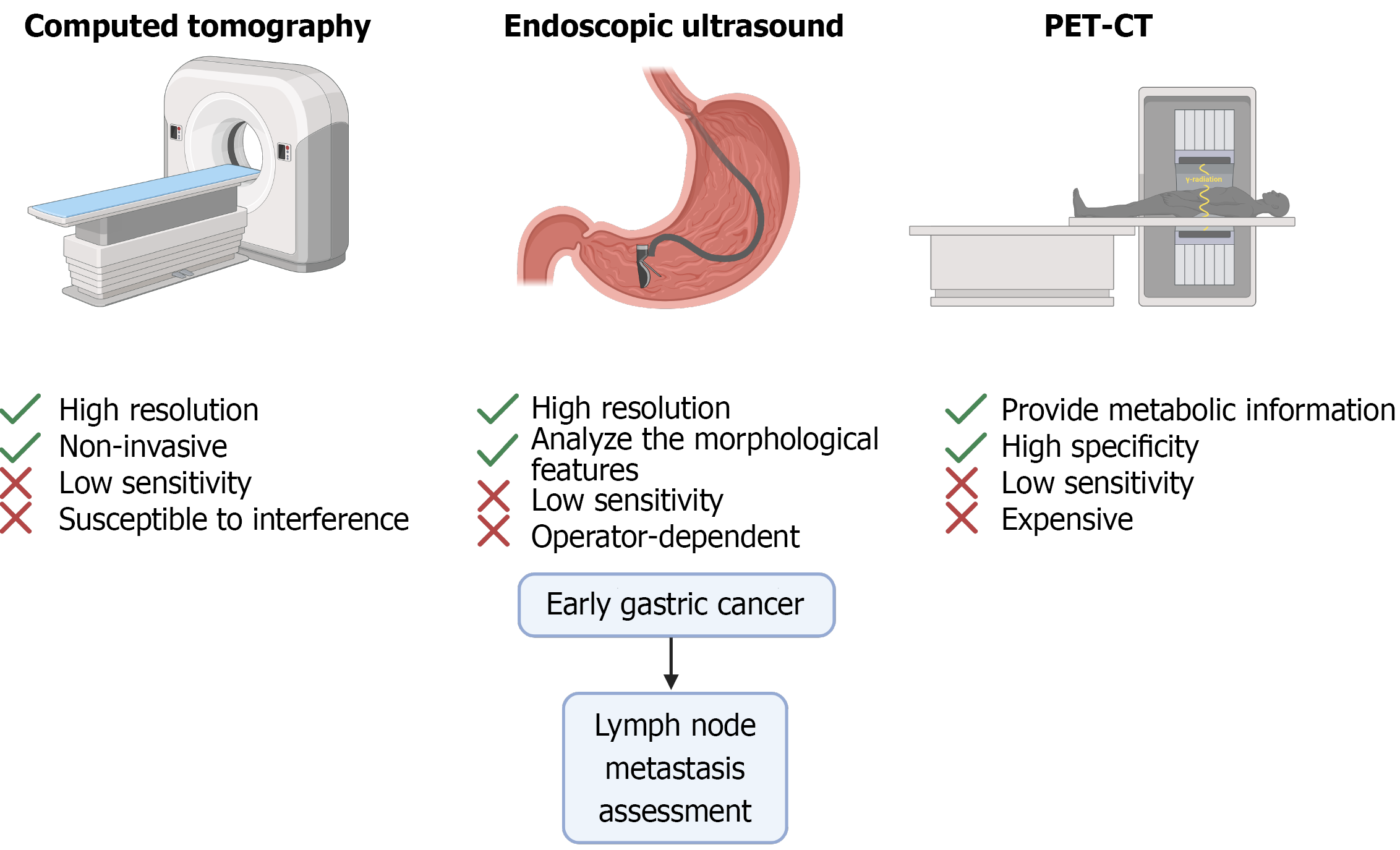
Figure 4 Comparative overview of imaging modalities for lymph node metastasis assessment in early gastric cancer.
Computed tomography, endoscopic ultrasound, and positron emission tomography-computed tomography provide complementary anatomical, morphological, and metabolic information, respectively. These modalities have distinct advantages and limitations and collectively contribute to preoperative evaluation of lymph node metastasis in early gastric cancer. PET-CT: Positron emission tomography-computed tomography.
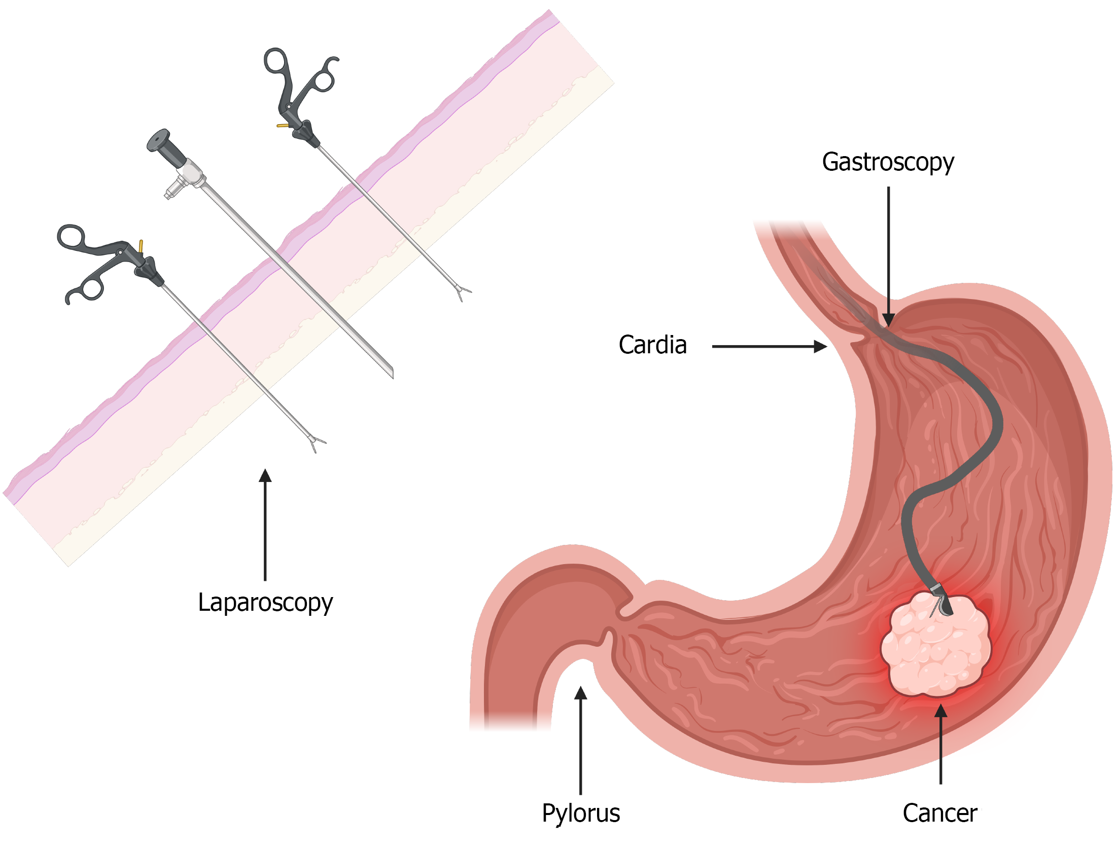
Figure 5 Laparoscopic-assisted endoscopic resection.
The endoscopic surgeon performs submucosal resection of early gastric cancer, while the laparoscopic team provides external assistance to optimize surgical exposure.
- Citation: Xia MT, Zhou Q, Han H, Zhang D, Fan X. Lymph node metastasis in early gastric cancer: A review of risk stratification and surgical decision-making. World J Gastrointest Oncol 2026; 18(10): 123261
- URL: https://www.wjgnet.com/1948-5204/full/v18/i10/123261.htm
- DOI: https://dx.doi.org/10.4251/wjgo.123261