Copyright
©The Author(s) 2025.
World J Gastrointest Oncol. May 15, 2025; 17(5): 104194
Published online May 15, 2025. doi: 10.4251/wjgo.v17.i5.104194
Published online May 15, 2025. doi: 10.4251/wjgo.v17.i5.104194
Figure 1 Ultrasound images show changes in the gastric wall, including thickening of the mucosal layer and narrowing of the gastric cavity.
A: The gastric cavity is well distended; B: With diffuse thickening of the mucosal layer in the antral; C: Body regions of the stomach, appearing rough and indistinct in local layers. The mucosal surface is uneven, with narrowing of the antral lumen.
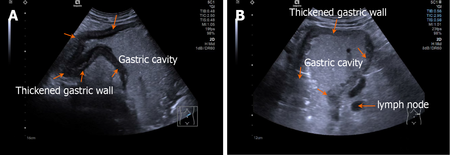
Figure 2 Ultrasonographic findings in gastric cancer.
A: The gastric wall layers are clearly defined, with circumferential thickening of the gastric wall in the antral and body regions, more pronounced in the antrum, narrowing of the antral lumen, an uneven mucosal surface, and partial interruption of the mucosa can be observed; B: Enlargement of the perigastric lymph nodes.
- Citation: Jiang Y, Xu SH, Han L, Lu N, Huang S, Wang L. Accuracy of dual-contrast gastrointestinal ultrasonography in predicting lymph node metastasis in older adults with gastric cancer. World J Gastrointest Oncol 2025; 17(5): 104194
- URL: https://www.wjgnet.com/1948-5204/full/v17/i5/104194.htm
- DOI: https://dx.doi.org/10.4251/wjgo.v17.i5.104194


