Copyright
©The Author(s) 2022.
World J Gastrointest Oncol. Dec 15, 2022; 14(12): 2329-2339
Published online Dec 15, 2022. doi: 10.4251/wjgo.v14.i12.2329
Published online Dec 15, 2022. doi: 10.4251/wjgo.v14.i12.2329
Figure 1 In vitro and in vivo cytotoxicity experiments of nucleus accumbens-1 on colon cancer cells.
A: The effect of nucleus accumbens-1 (NAC-1) on the toxicity of CD8+ T cells on the colon cancer cell line RKO; B: The effects of NAC-1, CD8+ T cells and MEK inhibitors on tumor killing in vivo (aP < 0.05). NAC-1: Nucleus accumbens-1.
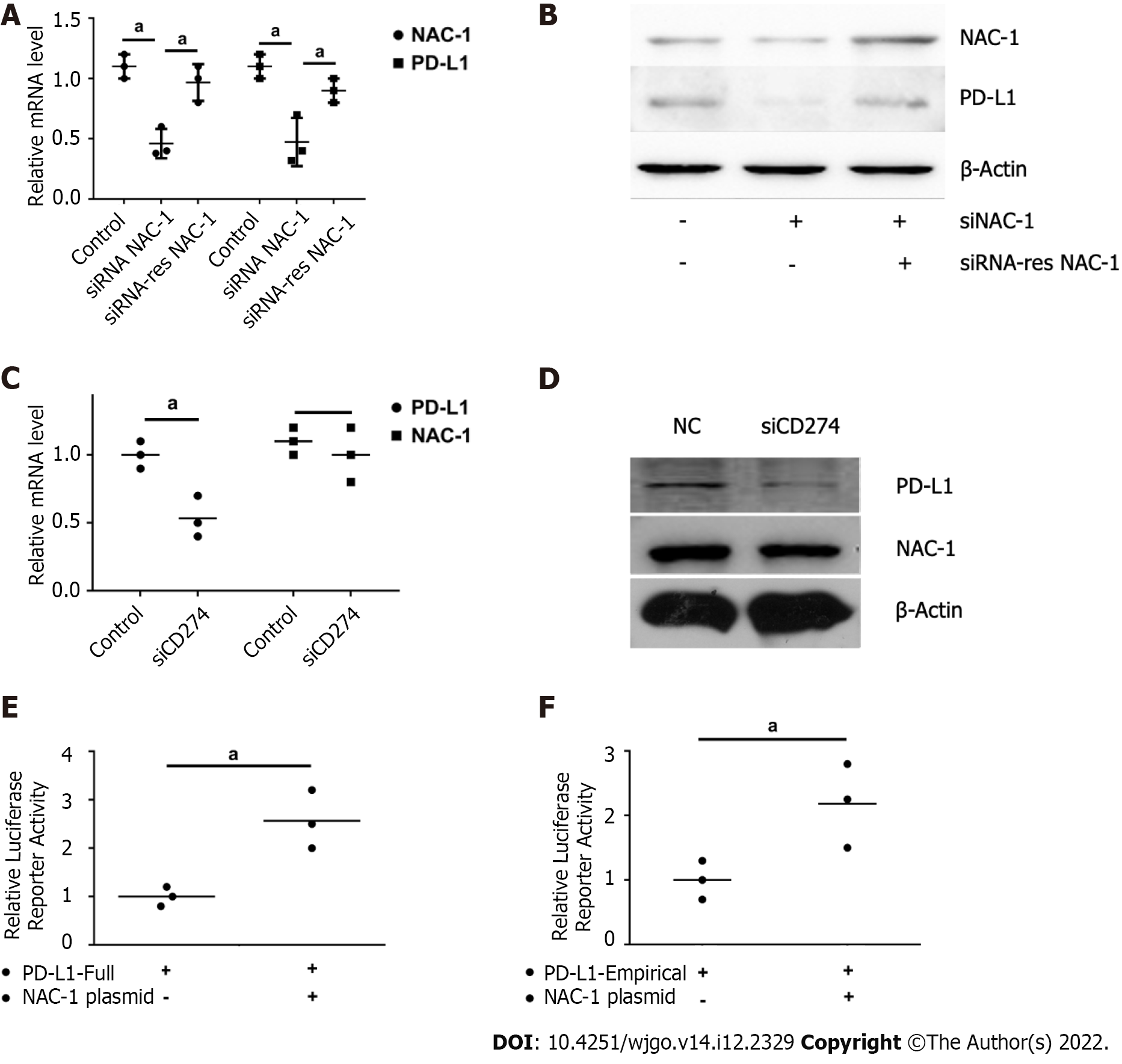
Figure 2 Effect of nucleus accumbens-1 on the expression of programmed death receptor-1 ligand.
A: Effect of nucleus accumbens-1 (NAC-1) on the mRNA expression of programmed death receptor-1 ligand (PD-L1); B: Effect of NAC-1 on the protein expression of PD-L1; C: The effect of PD-L1 on the transcription level of NAC-1; D: The effect of PD-L1 on the translation level of NAC-1; E: The effect of NAC-1 on the full promoter region of PD-L1; F: The effect of NAC-1 on the PD-L1 classic transcription factor binding segment (1:1, 2:1, 5:1, 10:1 are the ratios of CD8+ T cells to RKO; aP < 0.05). NAC-1: Nucleus accumbens-1; PD-L1: Programmed death receptor-1 ligand.
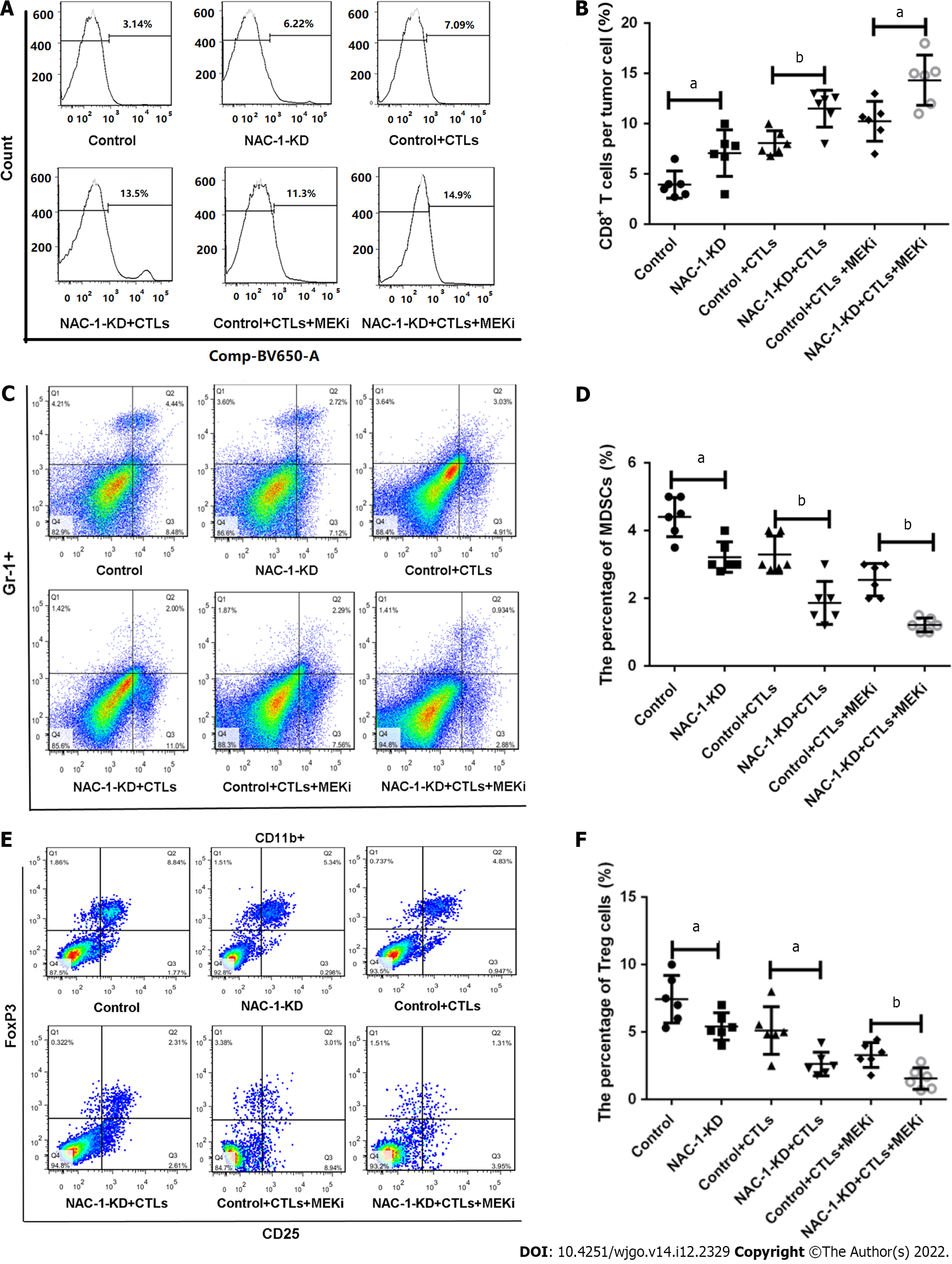
Figure 3 The effect of nucleus accumbens-1 on CD8+ T cells, myeloid-derived suppressor cells and regulatory T cells in the mouse colon cancer tumor microenvironment.
A: Flow cytometric detection of the effects of nucleus accumbens-1 (NAC-1), CD8+ T cells and MEK inhibitors on the expression of CD8+ T cells; B: Statistical analysis of CD8+ T-cell expression levels in each group; C: Flow cytometric detection of the effect of NAC-1 on the expression of myeloid-derived suppressor cells (MDSCs); D: Statistical analysis of the expression of MDSCs in each group; E: Flow cytometric detection of the effect of NAC-1 on the expression of regulatory T cell (Treg) cells; F: Statistical analysis of Treg expression in each group (aP < 0.05, bP < 0.01). NAC-1: Nucleus accumbens-1.
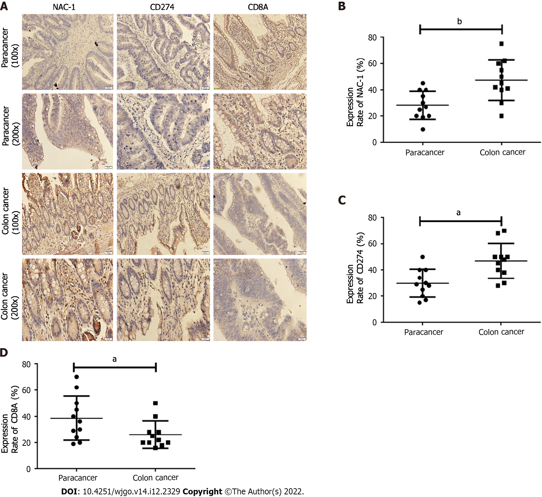
Figure 4 Exploring the relationship between nucleus accumbens-1, programmed death receptor-1 ligand and CD8 at the clinical level.
A: Nucleus accumbens-1 (NAC-1), programmed death receptor-1 ligand (PD-L1), and CD8 staining results of typical tissue samples from several patients, scale bar = 50 μm; B: NAC-1 expression in tumor tissues and adjacent tissues; C: PD-L1 expression in tumor tissues and adjacent tissues; D: The expression of CD8 in tumor tissues and adjacent tissues. NAC-1: Nucleus accumbens-1.
- Citation: Shen ZH, Luo WW, Ren XC, Wang XY, Yang JM. Expression of nucleus accumbens-1 in colon cancer negatively modulates antitumor immunity. World J Gastrointest Oncol 2022; 14(12): 2329-2339
- URL: https://www.wjgnet.com/1948-5204/full/v14/i12/2329.htm
- DOI: https://dx.doi.org/10.4251/wjgo.v14.i12.2329


