Copyright
©The Author(s) 2021.
World J Gastrointest Oncol. Apr 15, 2021; 13(4): 223-230
Published online Apr 15, 2021. doi: 10.4251/wjgo.v13.i4.223
Published online Apr 15, 2021. doi: 10.4251/wjgo.v13.i4.223
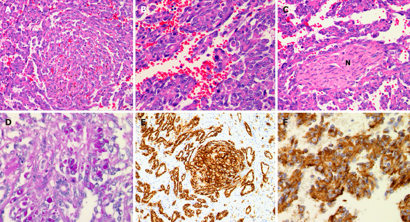
Figure 1 Histologic findings of pediatric hepatic angiosarcoma.
A: Whorls of atypical spindled cells (Hematoxylin-eosin staining, 10 ×); B: Moderate power view of the lesional cells show moderate nuclear atypia (Hematoxylin-eosin staining, 20 ×); C: Perineural invasion is be noted in this infiltrative lesion; note the nerve bundle (N) in the center (hematoxylin-eosin staining, 10 ×); D: Periodic acid-Schiff-positive eosinophilic globules are focally identified in the lesion, usually seen in association with the whorls of atypical spindled cells/glomeruloid bodies (para-aminosailcylic acid, 10 ×); E: The lesional cells are immunoreactive for vascular markers such as CD31 (10 ×) (note the glomeruloid body in the center); F: Factor VIII (20 ×).
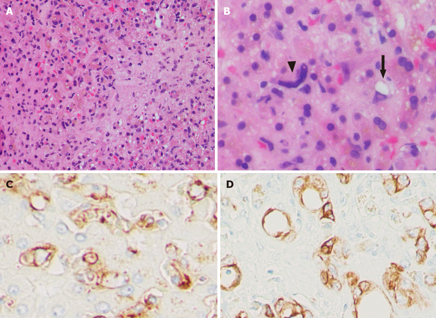
Figure 2 Pediatric hepatic epithelioid hemangioendothelioma.
A: Hypocellular epithelioid cells embedded in fibrotic stroma (right); entrapped hepatocytes with cholestasis are noted on the left (hematoxylin-eosin staining, 4 ×); B: High power view of the lesion demonstrates scattered atypical cells (arrowhead) with occasional intracytoplasmic lumen formation (arrow) (Hematoxylin-eosin staining, 20 ×); C: The neoplastic cells are immunoreactive for CD31 (20 ×); D: The neoplastic cells are immunoreactive for CK7 (20 ×).
- Citation: Bannoura S, Putra J. Primary malignant vascular tumors of the liver in children: Angiosarcoma and epithelioid hemangioendothelioma. World J Gastrointest Oncol 2021; 13(4): 223-230
- URL: https://www.wjgnet.com/1948-5204/full/v13/i4/223.htm
- DOI: https://dx.doi.org/10.4251/wjgo.v13.i4.223


