Copyright
©The Author(s) 2019.
World J Gastrointest Oncol. Jun 15, 2019; 11(6): 489-498
Published online Jun 15, 2019. doi: 10.4251/wjgo.v11.i6.489
Published online Jun 15, 2019. doi: 10.4251/wjgo.v11.i6.489
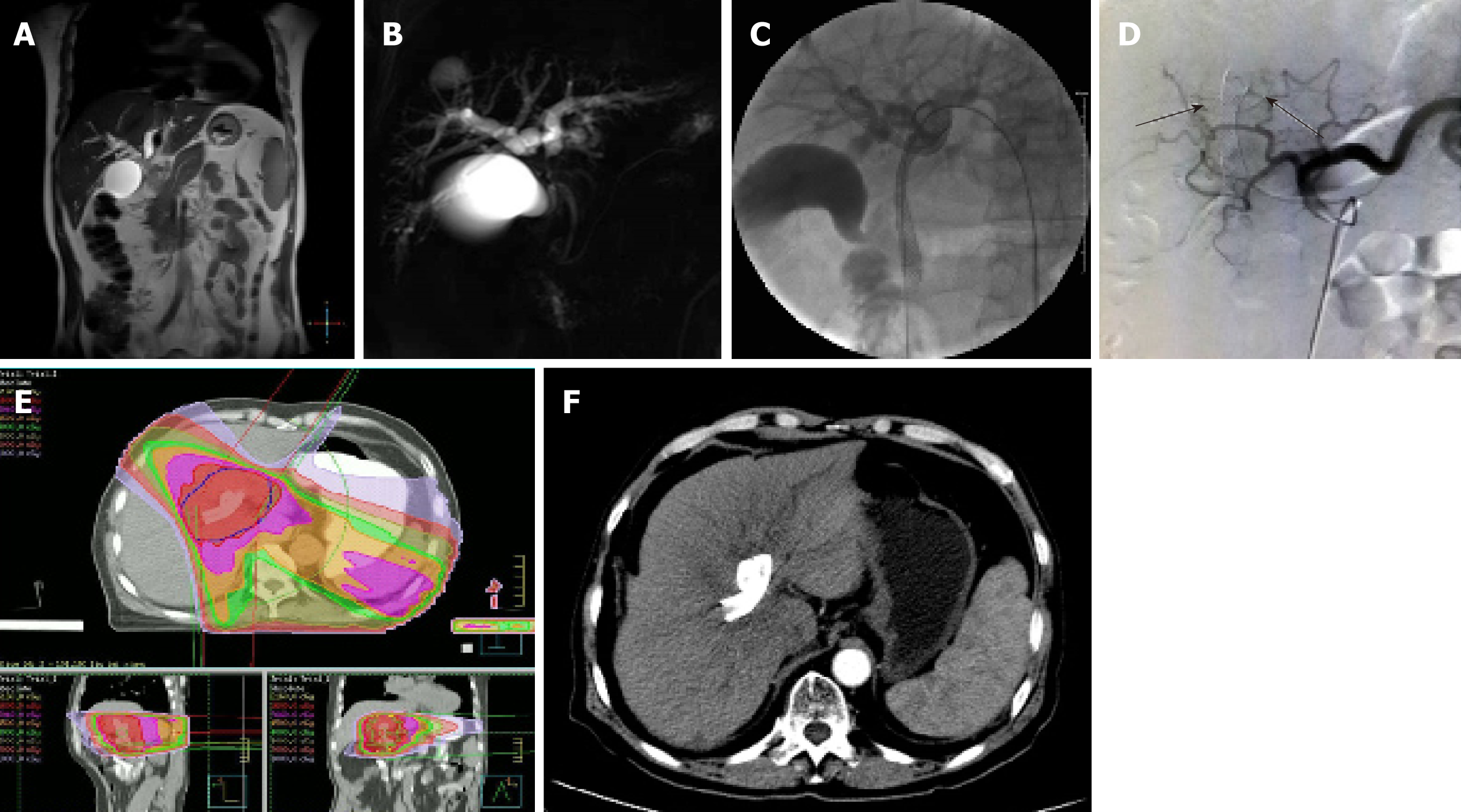
Figure 1 A 65-year-old man diagnosed with hilar cholangiocarcinoma.
A and B: Magnetic resonance imaging (A) and magnetic resonance cholangiopancreatography (B) showed left hepatic duct and right hepatic duct branch involvement (Bismuth-Corlette type IIIa); C: Patients underwent percutaneous double stent placement; D: During transcatheter arterial chemoembolization, the branches of the hepatic artery were responsible for the blood supply of the lesion area in the hepatic angiography (arrows); E: Intensity-modulated radiotherapy plan is shown; F: Liver cirrhosis was observed at 11 mo after receiving treatment.
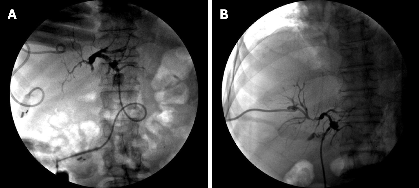
Figure 2 A patient with hilar cholangiocarcinoma who underwent biliary drainage.
A: Before transcatheter arterial chemoembolization combined with radiotherapy, the obstruction of the junction between the left and right hepatic ducts was shown during cholangiography; B: The obstruction was reduced and local stenosis was observed after treatment.
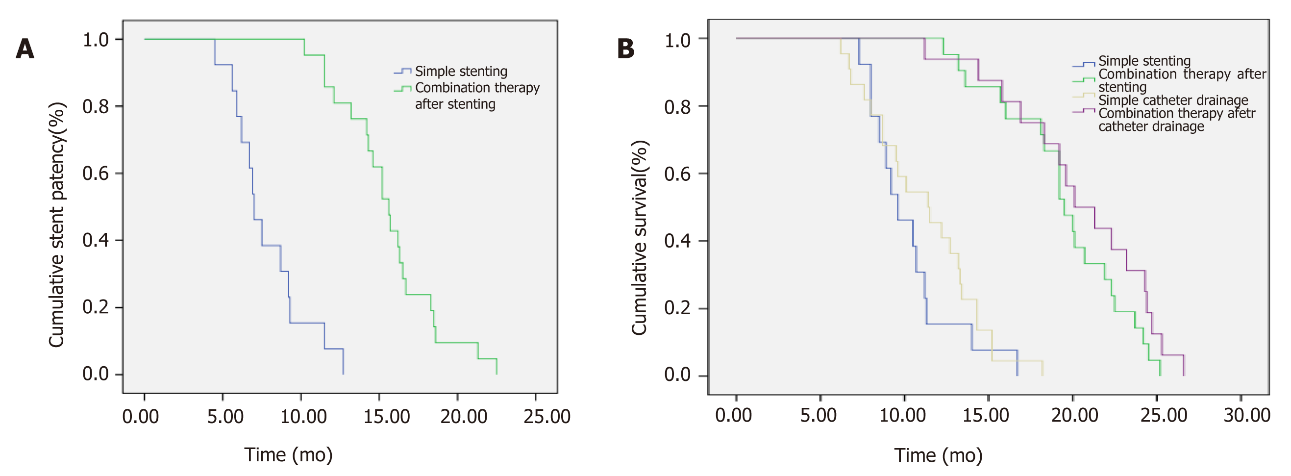
Figure 3 Comparison of stent patency time and survival between control group and combined treatment group.
A: Comparison of stent patency time; B: Comparison of survival.
- Citation: Zheng WH, Yu T, Luo YH, Wang Y, Liu YF, Hua XD, Lin J, Ma ZH, Ai FL, Wang TL. Clinical efficacy of gemcitabine and cisplatin-based transcatheter arterial chemoembolization combined with radiotherapy in hilar cholangiocarcinoma. World J Gastrointest Oncol 2019; 11(6): 489-498
- URL: https://www.wjgnet.com/1948-5204/full/v11/i6/489.htm
- DOI: https://dx.doi.org/10.4251/wjgo.v11.i6.489


