Copyright
©2012 Baishideng Publishing Group Co.
World J Gastrointest Endosc. Jul 16, 2012; 4(7): 335-338
Published online Jul 16, 2012. doi: 10.4253/wjge.v4.i7.335
Published online Jul 16, 2012. doi: 10.4253/wjge.v4.i7.335
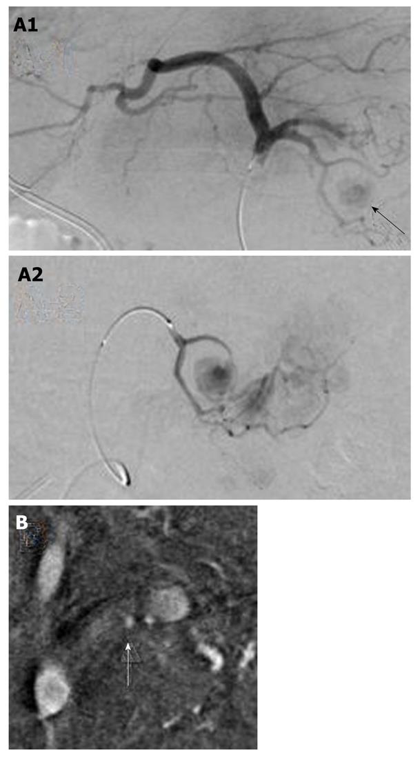
Figure 5 Angiography findings.
A1: The pseudoaneurysm of the left gastric artery was diagnosed on angiography (arrow). The left hepatic artery diverged from the left gastric artery; A2: The microcatheter was advanced in the region of the pseudoaneurysm, and the pseudoaneurysm was embolized with histoacryl and lipiodol; B: A small pseudoaneurysm was observed in the splenic artery (arrow), and the splenic artery was embolized by coils.
- Citation: Fukatsu K, Ueda K, Maeda H, Yamashita Y, Itonaga M, Mori Y, Moribata K, Shingaki N, Deguchi H, Enomoto S, Inoue I, Maekita T, Iguchi M, Tamai H, Kato J, Ichinose M. A case of chronic pancreatitis in which endoscopic ultrasonography was effective in the diagnosis of a pseudoaneurysm. World J Gastrointest Endosc 2012; 4(7): 335-338
- URL: https://www.wjgnet.com/1948-5190/full/v4/i7/335.htm
- DOI: https://dx.doi.org/10.4253/wjge.v4.i7.335


