Copyright
©The Author(s) 2015.
World J Gastrointest Endosc. Apr 16, 2015; 7(4): 346-353
Published online Apr 16, 2015. doi: 10.4253/wjge.v7.i4.346
Published online Apr 16, 2015. doi: 10.4253/wjge.v7.i4.346
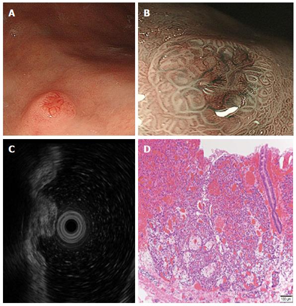
Figure 1 Type I gastric neuroendocrine tumor.
A: Conventional endoscopic image taken under white light. A hemispherical reddish polyp with a central depression is visible; B: Magnifying endoscopic image taken with a narrow band imaging system. Gastric pit-like structures present on the tumor’s surface, except for the central depression. In the central depression, the pit-like structure was not present, whereas dilated blackish-brown subepithelial vessels with cork-screw capillaries are visible; C: Endoscopic ultrasound showing a hypoechoic intramural structure in the second layer of the tumor; D: Histological appearance. Magnification (40 ×) of a hematoxylin-and-eosin–stained section of the tumor revealing a gastric neuroendocrine tumor limited to the mucosa.
- Citation: Sato Y. Endoscopic diagnosis and management of type I neuroendocrine tumors. World J Gastrointest Endosc 2015; 7(4): 346-353
- URL: https://www.wjgnet.com/1948-5190/full/v7/i4/346.htm
- DOI: https://dx.doi.org/10.4253/wjge.v7.i4.346


