Copyright
©2012 Baishideng Publishing Group Co.
World J Gastrointest Endosc. Mar 16, 2012; 4(3): 50-56
Published online Mar 16, 2012. doi: 10.4253/wjge.v4.i3.50
Published online Mar 16, 2012. doi: 10.4253/wjge.v4.i3.50
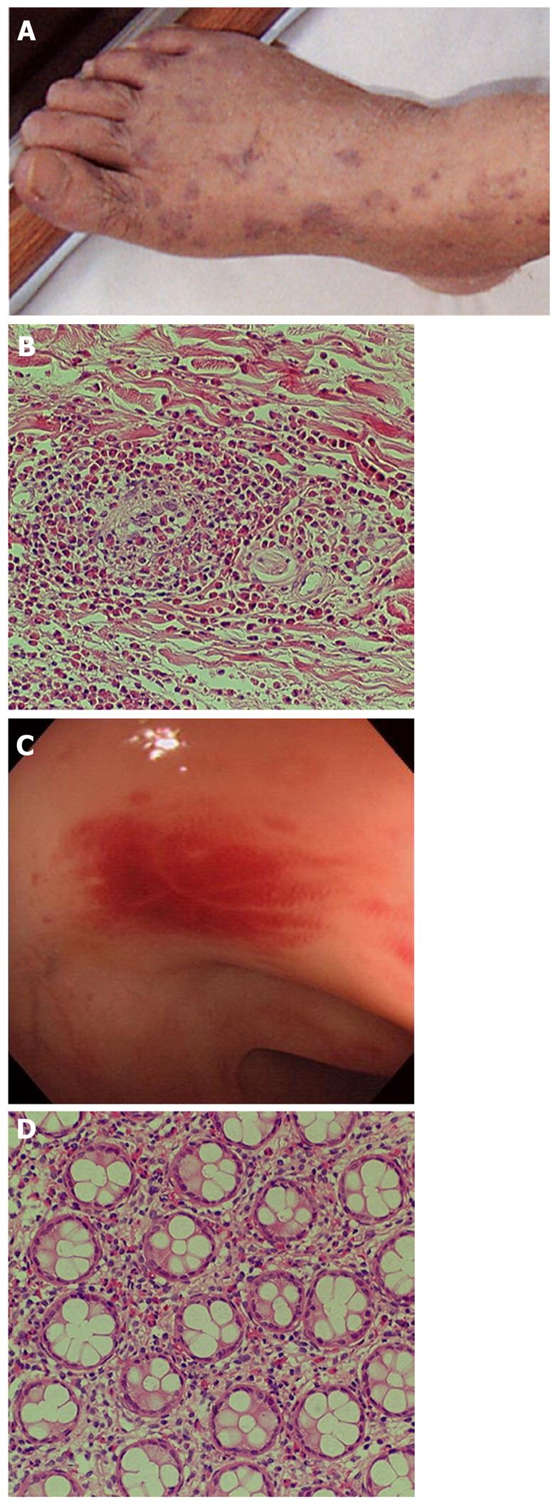
Figure 1 Churg-Strauss syndrome in a 60-year-old man with fever, abdominal pain, diarrhea, facial swelling, and purpura of the lower extremities.
A: Purpura of the right foot; B: Biopsy of the purpura revealed small vessel vasculitis with marked inflammatory infiltrate of eosinophils; C: Colonoscopy disclosed numerous areas of patchy mucosal erythema from the sigmoid colon to the splenic flexure; D: Biopsy of erythema showed mild infiltration of eosinophils around crypts. All figures and legends are reproduced from Hokama et al[11] with permission from Elsevier.
Figure 2 Henoch-Schönlein purpura in a 38-year-old man with hematochezia.
A: Palpable purpura of the right foot; B: Contrast-enhanced computed tomography scan of the abdomen showed diffuse thickening of the ileum (target sign) with mesenteric hypervascularity in a palisading pattern (comb sign), suggesting ischemic ileitis; C, D: Single balloon enteroscopy showed edematous petechiae with linear ulcers in the affected ileum. All figures and legends are reproduced from Hokama et al[15] with permission from BMJ Publishing Group Ltd.
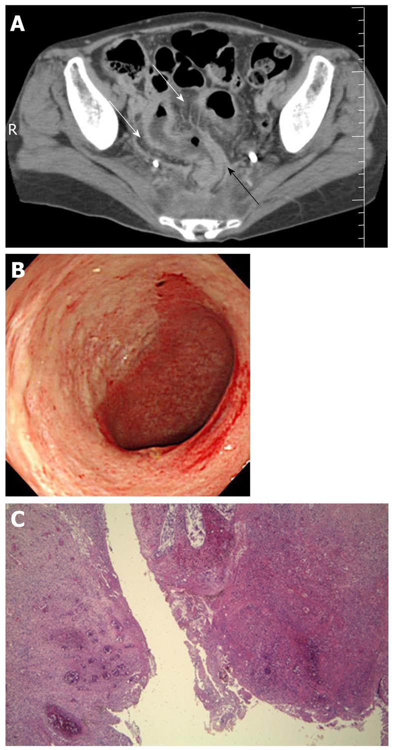
Figure 3 Systemic lupus erythematosus in a 40-year-old woman with lower abdominal pain and fever.
A: Contrast-enhanced computed tomography scan of the abdomen showed diffuse thickening of the rectosigmoid colon (black arrow) with engorgement of mesenteric vessels (comb sign, white arrows); B: Colonoscopy disclosed a large punched-out ulcer of the sigmoid colon; C: Perforation of the sigmoid colon occurred despite aggressive immunosuppressive therapy, requiring resection of the affected colon. The resected specimen disclosed bowel perforation with severe transmural inflammation, edema, hemorrhage and vasculitis (hematoxylin-eosin staining, ×40).
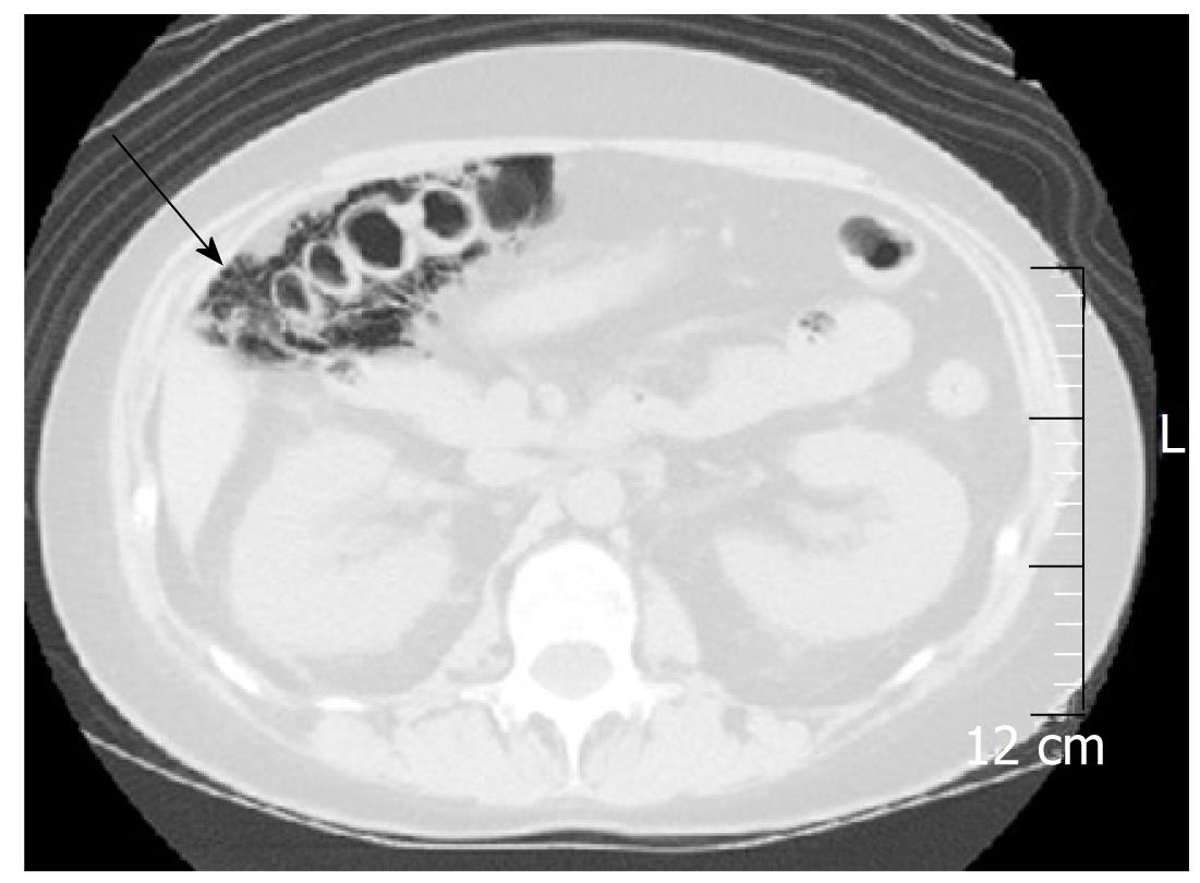
Figure 4 Systemic lupus erythematosus in a 23-year-old woman with abdominal pain and fever.
Plain computed tomography scan of the abdomen showed intramural gas of the ascending colon, suggesting pneumatosis intestinalis (arrow). Hyperbaric oxygen therapy was effective for improvement of the pneumatosis.
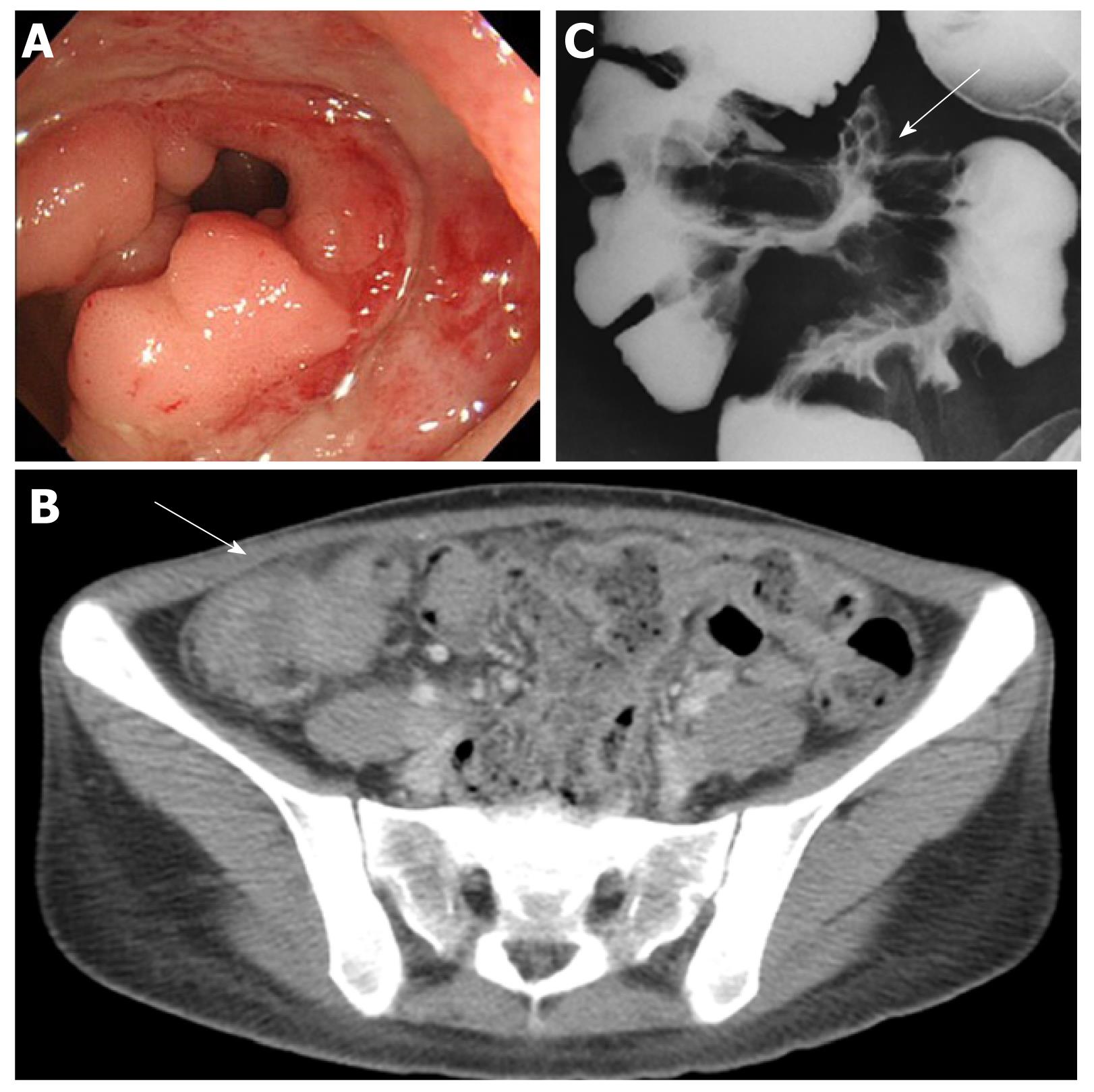
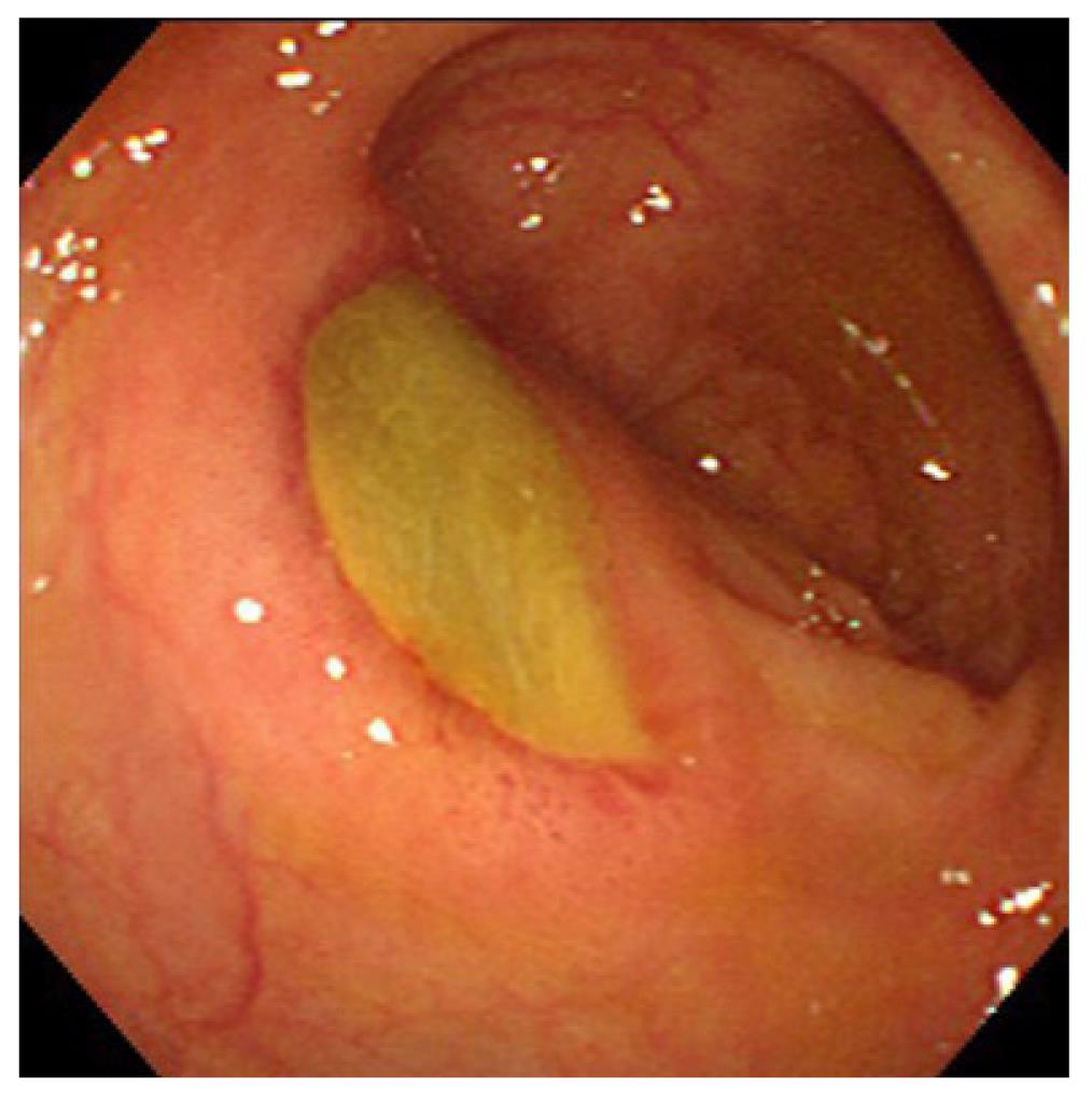
Figure 5 Behçet’s disease in a 25-year-old woman with abdominal pain and diarrhea.
A: Colonoscopy showed a large punched-out ulcer with elevated margins in the terminal ileum; B: Contrast-enhanced computed tomography scan of the abdomen showed a mass-like lesion with unevenly thickened bowel wall of the ileocecal region (arrow); C: Small bowel barium radiography disclosed the large ulcer (arrow) with convergence of mucosal folds in the terminal ileum.
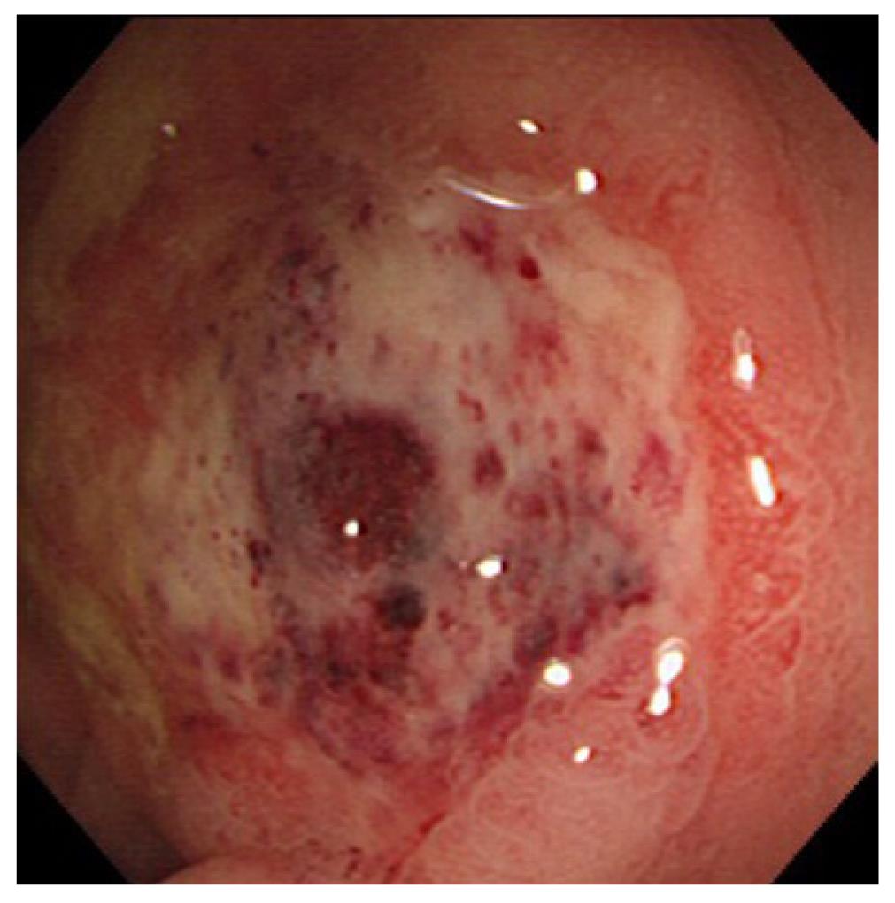
Figure 6 Behçet’s disease in a 50-year-old woman with abdominal pain and hematochezia-a large ovoid ulcer in the transverse colon.
The figure and legends are reproduced from Hokama et al[23] with permission from Elsevier.
Figure 7 Systemic lupus erythematosus in a 38-year-old woman with diarrhea.
Colonoscopy showed cytomegalovirus-associated round ulcer in the transverse colon.
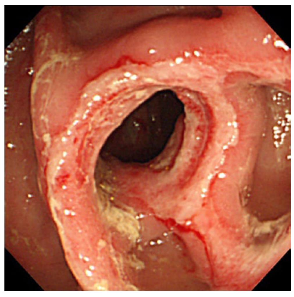
Figure 8 Rheumatoid arthritis in a 75-year-old woman with hematochezia.
Colonoscopy showed a diaphragm-like stricture with a circumferential ulcer in the rectum, making the diagnosis of non-steroidal anti-inflammatory drug-induced diaphragm disease. The figure and legends are reproduced from Hokama et al[33] with permission from BMJ Publishing Group Ltd.
- Citation: Hokama A, Kishimoto K, Ihama Y, Kobashigawa C, Nakamoto M, Hirata T, Kinjo N, Higa F, Tateyama M, Kinjo F, Iseki K, Kato S, Fujita J. Endoscopic and radiographic features of gastrointestinal involvement in vasculitis. World J Gastrointest Endosc 2012; 4(3): 50-56
- URL: https://www.wjgnet.com/1948-5190/full/v4/i3/50.htm
- DOI: https://dx.doi.org/10.4253/wjge.v4.i3.50


