Copyright
©2012 Baishideng Publishing Group Co.
World J Gastrointest Endosc. Mar 16, 2012; 4(3): 50-56
Published online Mar 16, 2012. doi: 10.4253/wjge.v4.i3.50
Published online Mar 16, 2012. doi: 10.4253/wjge.v4.i3.50
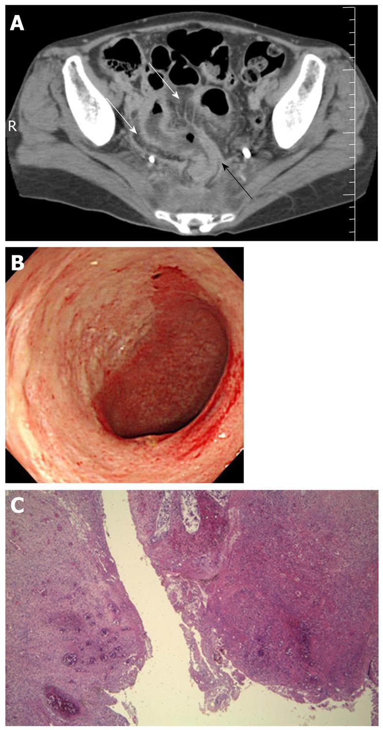
Figure 3 Systemic lupus erythematosus in a 40-year-old woman with lower abdominal pain and fever.
A: Contrast-enhanced computed tomography scan of the abdomen showed diffuse thickening of the rectosigmoid colon (black arrow) with engorgement of mesenteric vessels (comb sign, white arrows); B: Colonoscopy disclosed a large punched-out ulcer of the sigmoid colon; C: Perforation of the sigmoid colon occurred despite aggressive immunosuppressive therapy, requiring resection of the affected colon. The resected specimen disclosed bowel perforation with severe transmural inflammation, edema, hemorrhage and vasculitis (hematoxylin-eosin staining, ×40).
- Citation: Hokama A, Kishimoto K, Ihama Y, Kobashigawa C, Nakamoto M, Hirata T, Kinjo N, Higa F, Tateyama M, Kinjo F, Iseki K, Kato S, Fujita J. Endoscopic and radiographic features of gastrointestinal involvement in vasculitis. World J Gastrointest Endosc 2012; 4(3): 50-56
- URL: https://www.wjgnet.com/1948-5190/full/v4/i3/50.htm
- DOI: https://dx.doi.org/10.4253/wjge.v4.i3.50


