Copyright
©2010 Baishideng.
World J Gastrointest Endosc. Feb 16, 2010; 2(2): 54-60
Published online Feb 16, 2010. doi: 10.4253/wjge.v2.i2.54
Published online Feb 16, 2010. doi: 10.4253/wjge.v2.i2.54
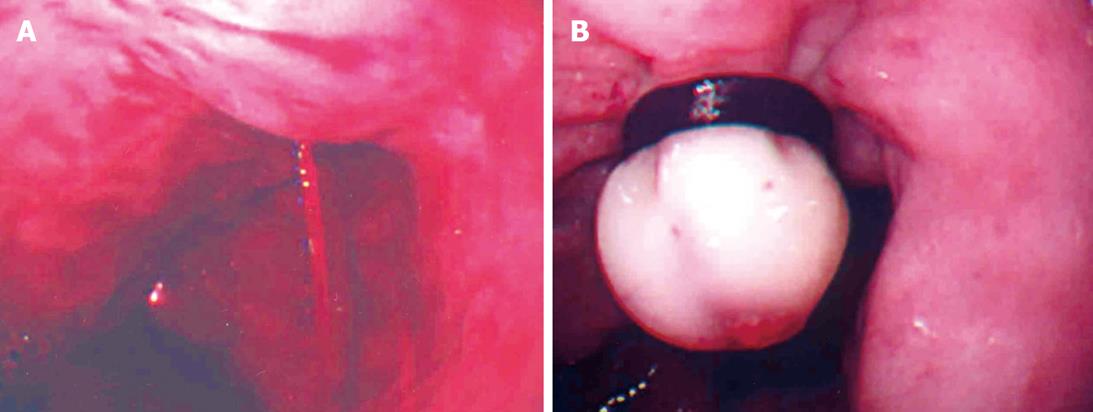
Figure 1 Use of EVL at the bleeding point.
A: A spurting bleeding from esophageal varices; B: Esophageal varices after EVL.
Figure 2 Changes of Esophageal varices after Endoscopic injection sclerotherapy.
A: Esophageal varices before an endoscopic therapy; B: Endoscopic injection sclerotherapy (EIS); C: Esophagus after EIS 2 years later.
Figure 3 Different patterns of bleeding of the gastric ulcer.
A: A spurting bleeding of the gastric ulcer (Forrest Ia); B oozing bleeding of the gastric ulcer (Forrest Ib); C: Non-bleeding visible vessel of the gastic ulcer (Forrest IIa).
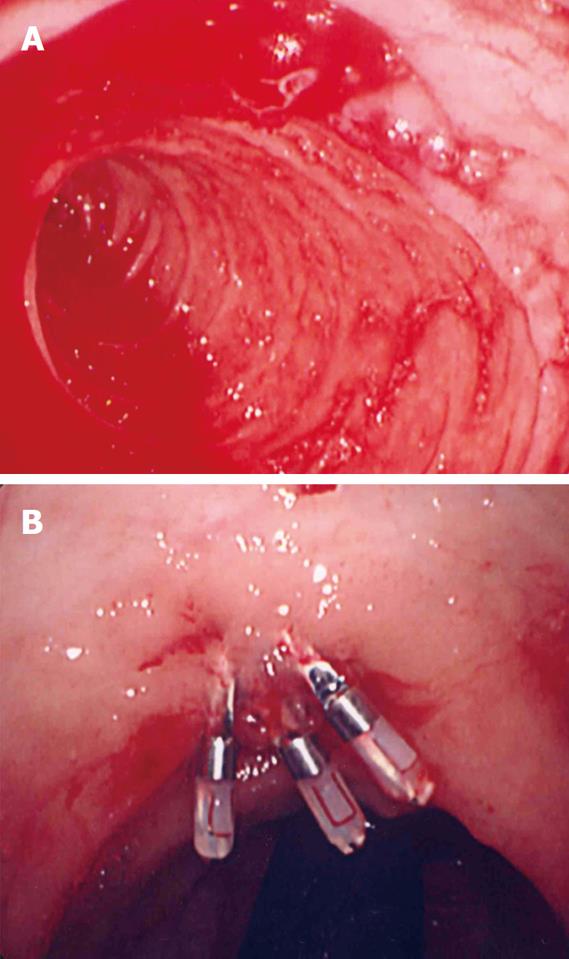
Figure 4 Duodenal diverticulum.
A: Hemorrhage; B: After endoscopic hemostasis.
- Citation: Anjiki H, Kamisawa T, Sanaka M, Ishii T, Kuyama Y. Endoscopic hemostasis techniques for upper gastrointestinal hemorrhage: A review. World J Gastrointest Endosc 2010; 2(2): 54-60
- URL: https://www.wjgnet.com/1948-5190/full/v2/i2/54.htm
- DOI: https://dx.doi.org/10.4253/wjge.v2.i2.54


