Copyright
©The Author(s) 2019.
World J Gastrointest Endosc. Mar 16, 2019; 11(3): 231-238
Published online Mar 16, 2019. doi: 10.4253/wjge.v11.i3.231
Published online Mar 16, 2019. doi: 10.4253/wjge.v11.i3.231
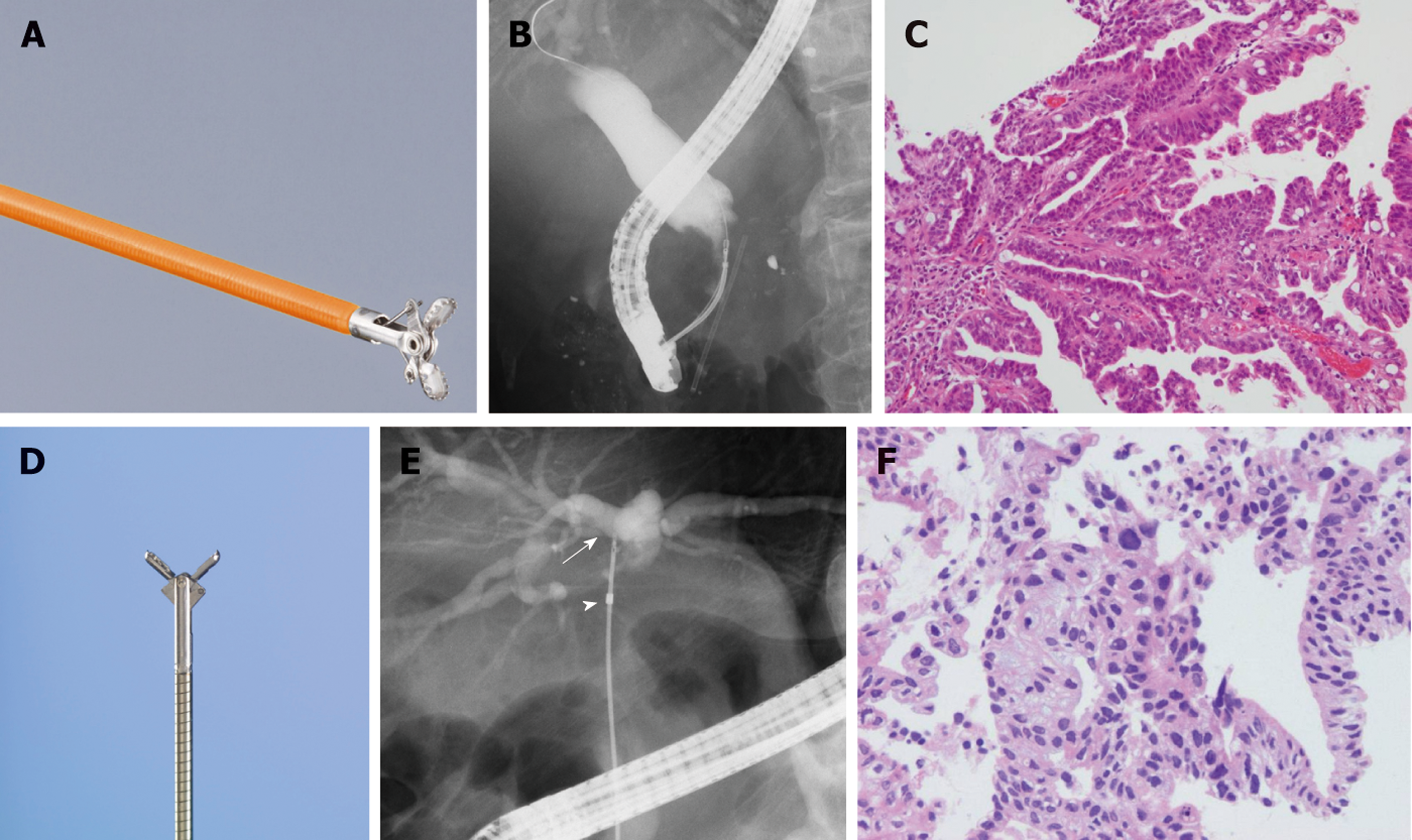
Figure 1 Biliary biopsy forceps and the procedural steps of biliary biopsies.
A: Radial JawTM 4 Biopsy Forceps. The cup diameter of these forceps is 2 mm. B: The image perspective in cholangiography. Biliary biopsy was performed with a Radial JawTM 4. C: The specimen was obtained with 2-mm biopsy forceps (X 200, hematoxylin eosin (HE) staining). Papillate lines were formed by biliary cancer cells. D: SpyBite. The cup diameter of these forceps is 1 mm. E: Biliary biopsy was performed with the SpyBite system through the MTW endoscopic retrograde cholangiopancreatography (ERCP) catheter (arrow: the tip of a SpyBite, arrowhead: the tip of an MTW ERCP catheter). F: The specimen was obtained by 1-mm biopsy forceps (X 400, HE stain). Papillate lines were formed by columnar biliary cancer cells.
- Citation: Takagi T, Sugimoto M, Suzuki R, Konno N, Asama H, Sato Y, Irie H, Watanabe K, Nakamura J, Kikuchi H, Takasumi M, Hashimoto M, Hikichi T, Ohira H. Appropriate number of biliary biopsies and endoscopic retrograde cholangiopancreatography sessions for diagnosing biliary tract cancer. World J Gastrointest Endosc 2019; 11(3): 231-238
- URL: https://www.wjgnet.com/1948-5190/full/v11/i3/231.htm
- DOI: https://dx.doi.org/10.4253/wjge.v11.i3.231


