Copyright
©2014 Baishideng Publishing Group Inc.
World J Hepatol. Jun 27, 2014; 6(6): 426-434
Published online Jun 27, 2014. doi: 10.4254/wjh.v6.i6.426
Published online Jun 27, 2014. doi: 10.4254/wjh.v6.i6.426
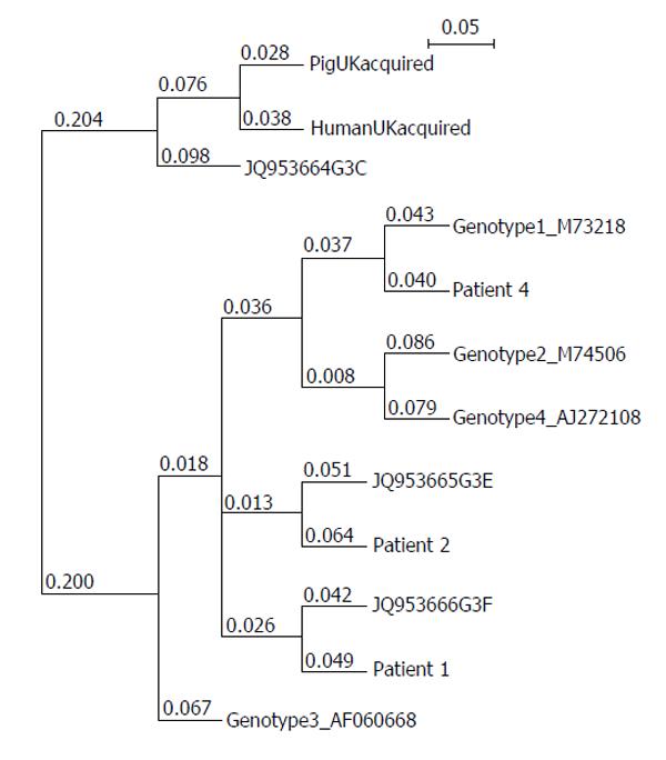
Figure 1 Phylogenetic relationship between the 4 hepatitis E virus genotype reference sequences and those isolated from a United Kingdom swine (AF503512), a United Kingdom patient with locally acquired hepatitis E virus (AY362357), genotype 3 subtypes described in[19] and the patients discussed in this paper.
Sequences were assembled using ClustalW and the phylogenetic tree expressed in the Newick format using NJ plot[20]. The 121 bp sequences correspond to nucleotides 6332-6476 of hepatitis E virus genotype 3 reference strain AF060668.
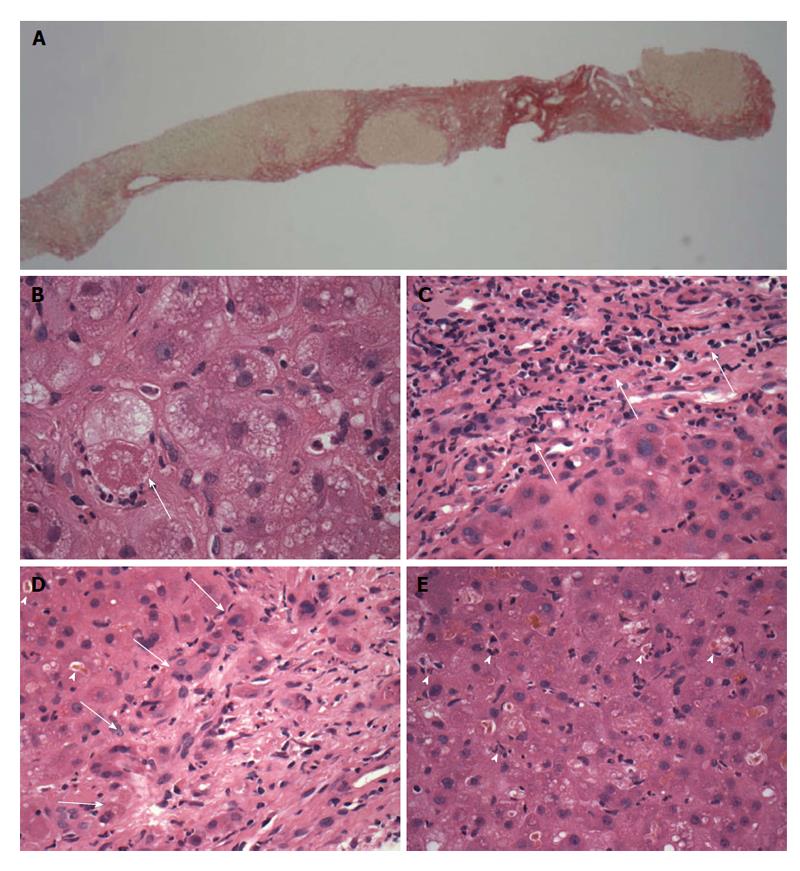
Figure 2 Patient 2, transjugular needle biopsy.
A: Low magnification view of part of the biopsy, showing red-stained nodular fibrosis indicating cirrhosis (x 20 original magnification, picrosirius red stain); B: Ballooned hepatocyte (arrow) containing a Mallory-Denk body and with surrounding neutrophils (satellitosis), features of steatohepatitis. (× 600 original magnification, H and E stain). Small clusters of these cells were present in the biopsy, with sparse small droplet macro-steatosis; C: Low grade hepatitic infiltrate (arrows) of lymphocytes with occasional plasma cells in the portal area (× 400 original magnification, H and E stain); D: Prominent cholangiolitis (periportal ductules with oedema and neutrophils) (region indicated by arrows), with adjacent liver parenchyma showing canalicular cholestasis (arrowheads); E: Lobule showing mild disarray with cholestasis, increased lymphocytes and Kupffer cells within sinusoids and scattered apoptotic/necrotic cells (arrowheads).
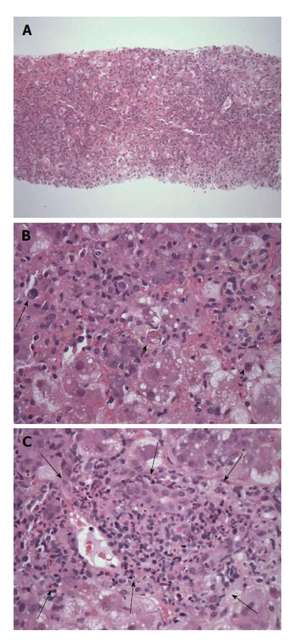
Figure 3 Patient 4, transjugular needle biopsy showing severe acute lobular hepatitis.
A: Low magnification view showing the diffuse nature of the liver inflammation and injury (original magnification × 40, H and E); B: Severely inflamed lobule with numerous infiltrating inflammatory cells, including occasional plasma cells (long arrow), hepatocyte cell death (short arrow) and ballooning injury (arrowhead) (original magnification × 400, H and E); C: Shows an inflamed portal tract (delineated by arrows) expanded within by mononuclear inflammatory cells without bile duct injury and with only rare plasma cells or eosinophils. There is neutrophil cholangiolitis around the portal tract (just within the arrows) but no prominent interface hepatitis (original magnification × 200, H and E).
- Citation: Crossan CL, Simpson KJ, Craig DG, Bellamy C, Davidson J, Dalton HR, Scobie L. Hepatitis E virus in patients with acute severe liver injury. World J Hepatol 2014; 6(6): 426-434
- URL: https://www.wjgnet.com/1948-5182/full/v6/i6/426.htm
- DOI: https://dx.doi.org/10.4254/wjh.v6.i6.426


