Copyright
©2014 Baishideng Publishing Group Inc.
World J Hepatol. Jun 27, 2014; 6(6): 426-434
Published online Jun 27, 2014. doi: 10.4254/wjh.v6.i6.426
Published online Jun 27, 2014. doi: 10.4254/wjh.v6.i6.426
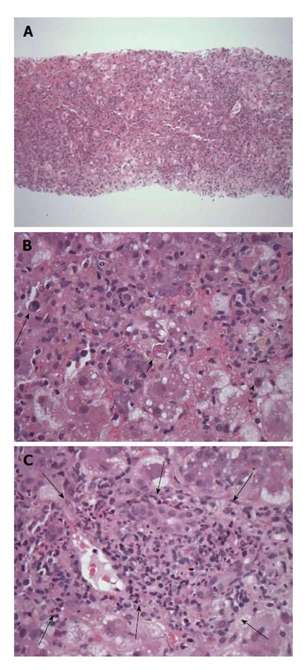
Figure 3 Patient 4, transjugular needle biopsy showing severe acute lobular hepatitis.
A: Low magnification view showing the diffuse nature of the liver inflammation and injury (original magnification × 40, H and E); B: Severely inflamed lobule with numerous infiltrating inflammatory cells, including occasional plasma cells (long arrow), hepatocyte cell death (short arrow) and ballooning injury (arrowhead) (original magnification × 400, H and E); C: Shows an inflamed portal tract (delineated by arrows) expanded within by mononuclear inflammatory cells without bile duct injury and with only rare plasma cells or eosinophils. There is neutrophil cholangiolitis around the portal tract (just within the arrows) but no prominent interface hepatitis (original magnification × 200, H and E).
- Citation: Crossan CL, Simpson KJ, Craig DG, Bellamy C, Davidson J, Dalton HR, Scobie L. Hepatitis E virus in patients with acute severe liver injury. World J Hepatol 2014; 6(6): 426-434
- URL: https://www.wjgnet.com/1948-5182/full/v6/i6/426.htm
- DOI: https://dx.doi.org/10.4254/wjh.v6.i6.426


