Copyright
©2014 Baishideng Publishing Group Inc.
World J Hepatol. Jun 27, 2014; 6(6): 426-434
Published online Jun 27, 2014. doi: 10.4254/wjh.v6.i6.426
Published online Jun 27, 2014. doi: 10.4254/wjh.v6.i6.426
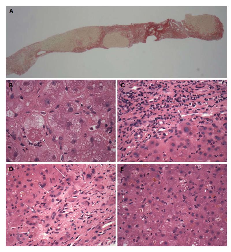
Figure 2 Patient 2, transjugular needle biopsy.
A: Low magnification view of part of the biopsy, showing red-stained nodular fibrosis indicating cirrhosis (x 20 original magnification, picrosirius red stain); B: Ballooned hepatocyte (arrow) containing a Mallory-Denk body and with surrounding neutrophils (satellitosis), features of steatohepatitis. (× 600 original magnification, H and E stain). Small clusters of these cells were present in the biopsy, with sparse small droplet macro-steatosis; C: Low grade hepatitic infiltrate (arrows) of lymphocytes with occasional plasma cells in the portal area (× 400 original magnification, H and E stain); D: Prominent cholangiolitis (periportal ductules with oedema and neutrophils) (region indicated by arrows), with adjacent liver parenchyma showing canalicular cholestasis (arrowheads); E: Lobule showing mild disarray with cholestasis, increased lymphocytes and Kupffer cells within sinusoids and scattered apoptotic/necrotic cells (arrowheads).
- Citation: Crossan CL, Simpson KJ, Craig DG, Bellamy C, Davidson J, Dalton HR, Scobie L. Hepatitis E virus in patients with acute severe liver injury. World J Hepatol 2014; 6(6): 426-434
- URL: https://www.wjgnet.com/1948-5182/full/v6/i6/426.htm
- DOI: https://dx.doi.org/10.4254/wjh.v6.i6.426


