Copyright
©2014 Baishideng Publishing Group Inc.
World J Hepatol. Dec 27, 2014; 6(12): 894-900
Published online Dec 27, 2014. doi: 10.4254/wjh.v6.i12.894
Published online Dec 27, 2014. doi: 10.4254/wjh.v6.i12.894
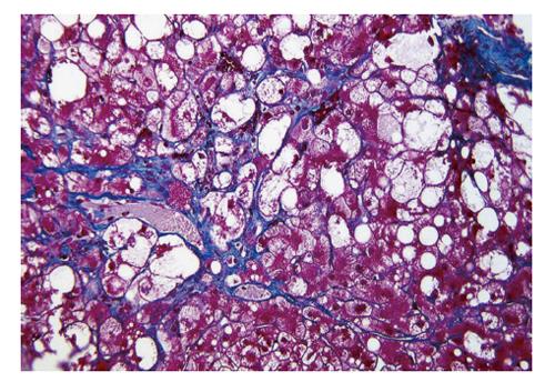
Figure 1 Typical case of nonalcoholic steatohepatitis showing hepatocellular ballooning and perivenular/pericellular fibrosis (Azan-Mallory stain; Original magnification, × 200).
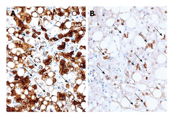
Figure 2 Typical immnohistochemical findings of nonalcoholic steatohepatitis.
A: Cytokeratin (CK)18; B: Ubiquitin. Stained (brown) small aggregates are seen in ballooned hepatocytes with CK18-negative cytoplasms (arrows) [Immunoperoxidase; original magnifications, (A) × 150 and (B) × 200].
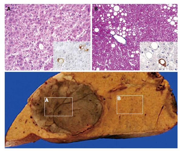
Figure 3 Case of hepatocellular carcinoma (A) associated with simple steatosis (B)[59] [upper row, histology with immunoperoxidase for a peroxidation marker (inset), original magnifications, (A) × 100 and (B) × 100; lower row, a macroscopic photo of the sample].
- Citation: Ikura Y. Transitions of histopathologic criteria for diagnosis of nonalcoholic fatty liver disease during the last three decades. World J Hepatol 2014; 6(12): 894-900
- URL: https://www.wjgnet.com/1948-5182/full/v6/i12/894.htm
- DOI: https://dx.doi.org/10.4254/wjh.v6.i12.894


