Copyright
©2010 Baishideng.
World J Hepatol. Jul 27, 2010; 2(7): 292-294
Published online Jul 27, 2010. doi: 10.4254/wjh.v2.i7.292
Published online Jul 27, 2010. doi: 10.4254/wjh.v2.i7.292
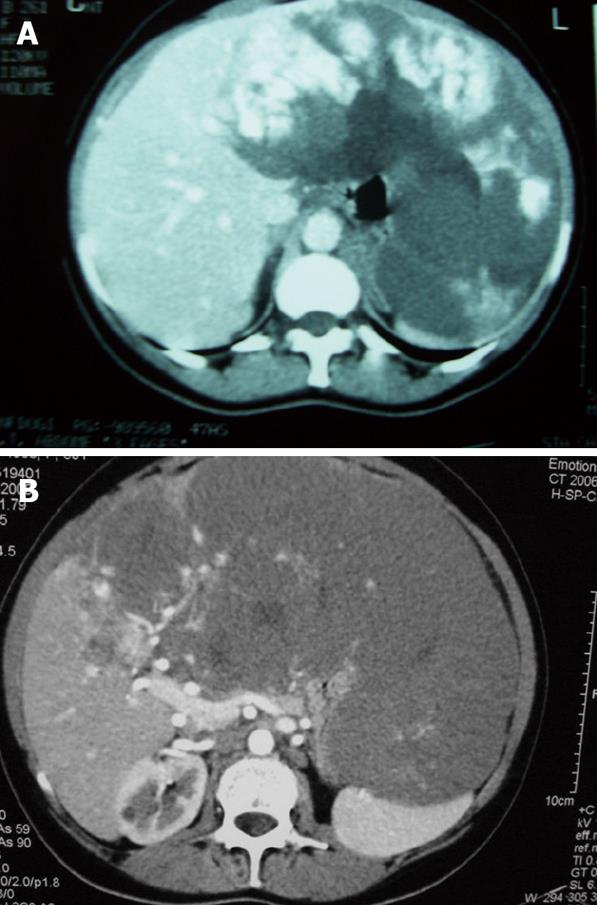
Figure 1 Computed tomography scan of a patients with a liver hemangioma.
A: The first scan performed 7 years ago, showing an oversized hemangioma on the left hepatic lobe; B: During the symptoms worsening a second CT-Scan showed tumor growth. The lesion reached the central portions of the liver.
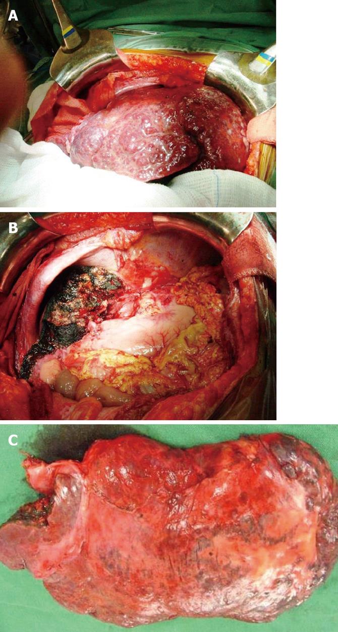
Figure 2 Surgical treatment of the patient with a liver hemangioma.
A: After the laparotomy, a giant hemangioma was identified, and this lesion led to a great anatomical deformity in the liver; B: After the resection, the remnant liver in the cleared abdominal cavity; C: The 40 cm-giant liver hemangioma was sent to anatomical and pathological analysis.
- Citation: Koszka AJ, Ferreira FG, Aquino CG, Ribeiro MA, Gallo AS, Aranzana EM, Szutan LA. Resection of a rapid-growing 40-cm giant liver hemangioma. World J Hepatol 2010; 2(7): 292-294
- URL: https://www.wjgnet.com/1948-5182/full/v2/i7/292.htm
- DOI: https://dx.doi.org/10.4254/wjh.v2.i7.292


