Copyright
©The Author(s) 2021.
World J Stem Cells. Jul 26, 2021; 13(7): 825-840
Published online Jul 26, 2021. doi: 10.4252/wjsc.v13.i7.825
Published online Jul 26, 2021. doi: 10.4252/wjsc.v13.i7.825
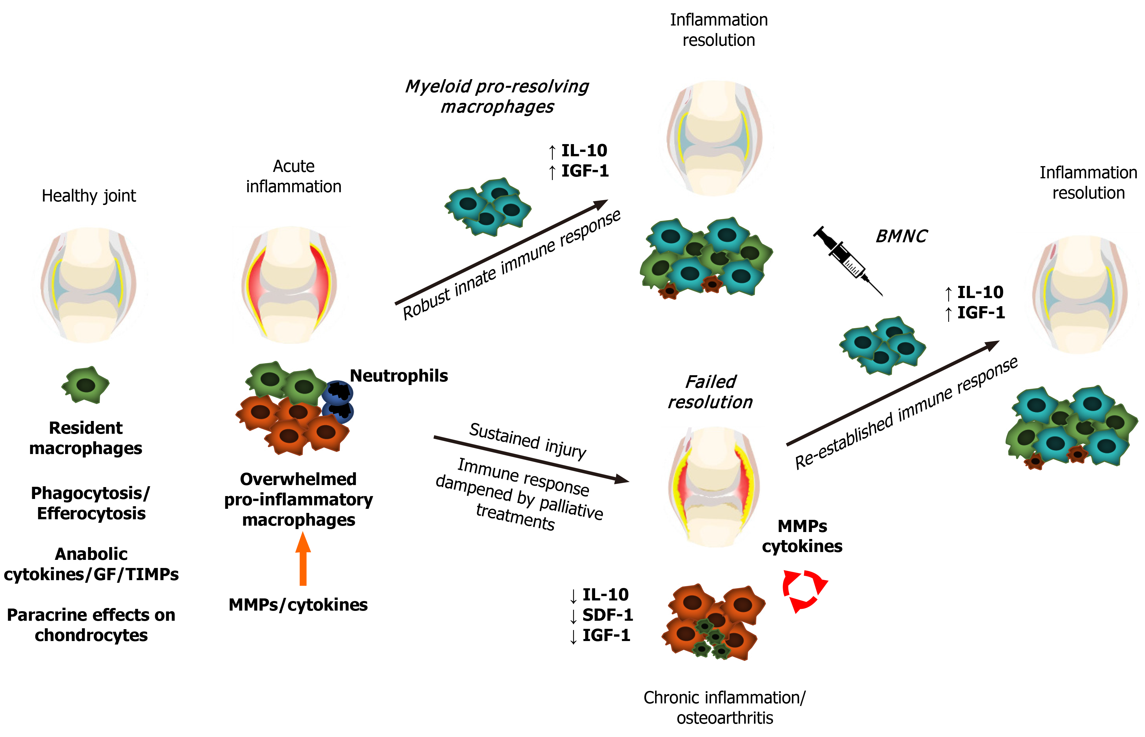
Figure 1 Schematic representation of macrophage status and responses during joint health and disease.
Macrophages promote synovial health through phagocytosis, efferocytosis, secretion of synovial fluid cytokines and growth factors, and paracrine effects on chondrocyte metabolism. Following damage, synovial macrophages form a shield to the injured site to further drive tissue repair. If the extent of the injury or continuing trauma overwhelms the shielding and repairing capacity of macrophages, macrophages increase expression of cytokines, including interleukin (IL)-1, IL-6, and tumor necrosis factor -α in a phlogistic inflammatory response, recruiting first neutrophils, followed by myeloid macrophages. These macrophages have a strong pro-resolving capacity associated with high production of IL-10 and insulin-like growth factor 1, which then leads to inflammation resolution and tissue repair. If interfering factors impair the resolution of the inflammatory process, chronic low-grade inflammation persists, leading to aberrant remodeling of synovial tissues and joint degeneration. Joint injection with bone marrow mononuclear cells in chronically inflamed joints augments the macrophage-mediated mechanisms of joint homeostasis, resolving joint inflammation. GF: Growth factors; TIMPs: Tissue inhibitors of metalloproteinases; MMPs: Matrix metalloproteinases; IL-10: Interleukin 10; IGF-1: Insulin-like growth factor 1; SDF-1: Stromal-derived factor 1; BMNC: Bone marrow mononuclear cells.
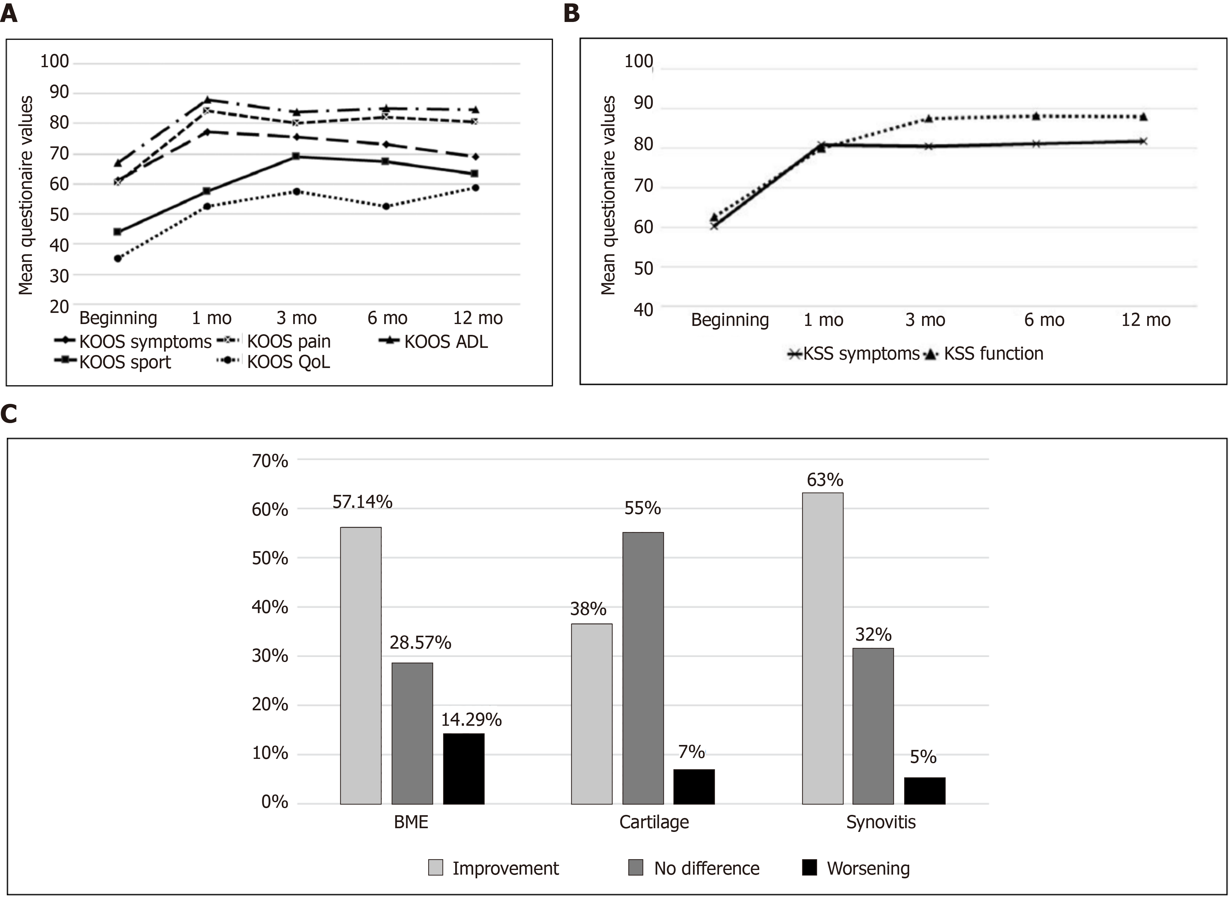
Figure 2 Clinical (Knee injury and osteoarthritis outcome score and Knee society score, n = 34) and imaging (magnetic resonance imaging, n = 30) outcomes from patients with knee Osteoarthritis treated with bone marrow mononuclear cells over 12 mo post-treatment[102].
A and B: Significant improvements (P < 0.05) were observed for Knee injury and osteoarthritis outcome score’ (A) and Knee Society Score (B) scores during the 12-mo follow-up period and most were sustained over time; C: Changes in bone marrow edema, cartilage and synovitis detected by magnetic resonance imaging 6 mo after treatment with bone marrow mononuclear cells. Citation: Goncars V, Kalnberzs K, Jakobsons E, Enģele I, Briede I, Blums K, Erglis K, Erglis M, Patetko L, Muiznieks I, Erglis A. Treatment of Knee Osteoarthritis with Bone Marrow–Derived Mononuclear Cell Injection: 12-Month Follow-up. Cartilage 2019; 10 (1): 26-35. Copyright © The Author(s) 2018. Published by SAGE Publications. KOOS: Knee injury and osteoarthritis outcome Score; KSS: Knee society score; QoL: Quality of life; ADL: Activities of daily living; BME: Bone marrow edema.
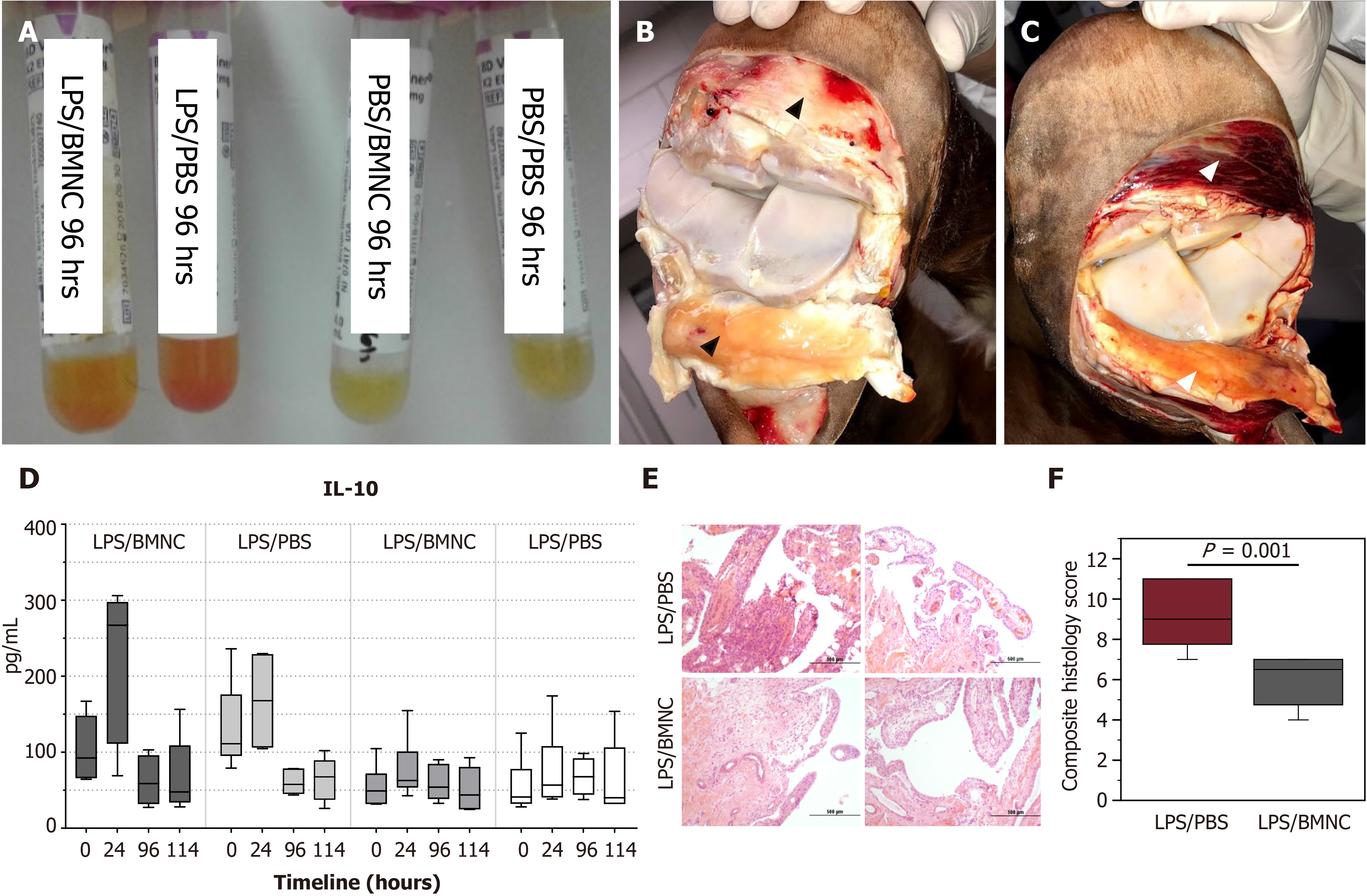
Figure 3 Gross and analytical findings from normal and inflamed equine joints treated with bone marrow mononuclear cells[55].
A: Improvements in synovial fluid associated with decreased cellularity and red cell contamination at 96 h compared to Phosphate buffered saline (PBS)-treated controls; B: Improvements in the synovium were reflected by decreased intra-and peri- and articular synovial hemorrhage and edema, in bone marrow mononuclear cells (BMNC)-(black arrowheads) compared to; C: PBS-treated joints (white arrowheads); D: There was a marked increase in synovial fluid interleukin 10 concentrations at 24 h in inflamed joints, which was much higher in BMNC-treated joints as compared with PBS-treated joints; E and F: BMNC-treated joints exhibited lower scores for all histological aspects of inflammation, although these were only significant (P < 0.05) for vascularity and the composite score(F); Citation: Menarim BC, Gillis KH, Oliver A, Mason C, Ngo Y, Were SR, Barrett SH, Luo X, Byron CR, Dahlgren. Autologous bone marrow mononuclear cells modulate joint homeostasis in an equine in vivo model of synovitis. FASEB J 33, 14337–14353. Copyright © The Author(s) 2019. Published by Wiley Online Library in cooperation with the Federation of American Societies for Experimental Biology. LPS: Lipopolysaccharide; PBS: Phosphate buffered saline; BMNC: Bone marrow mononuclear cells.
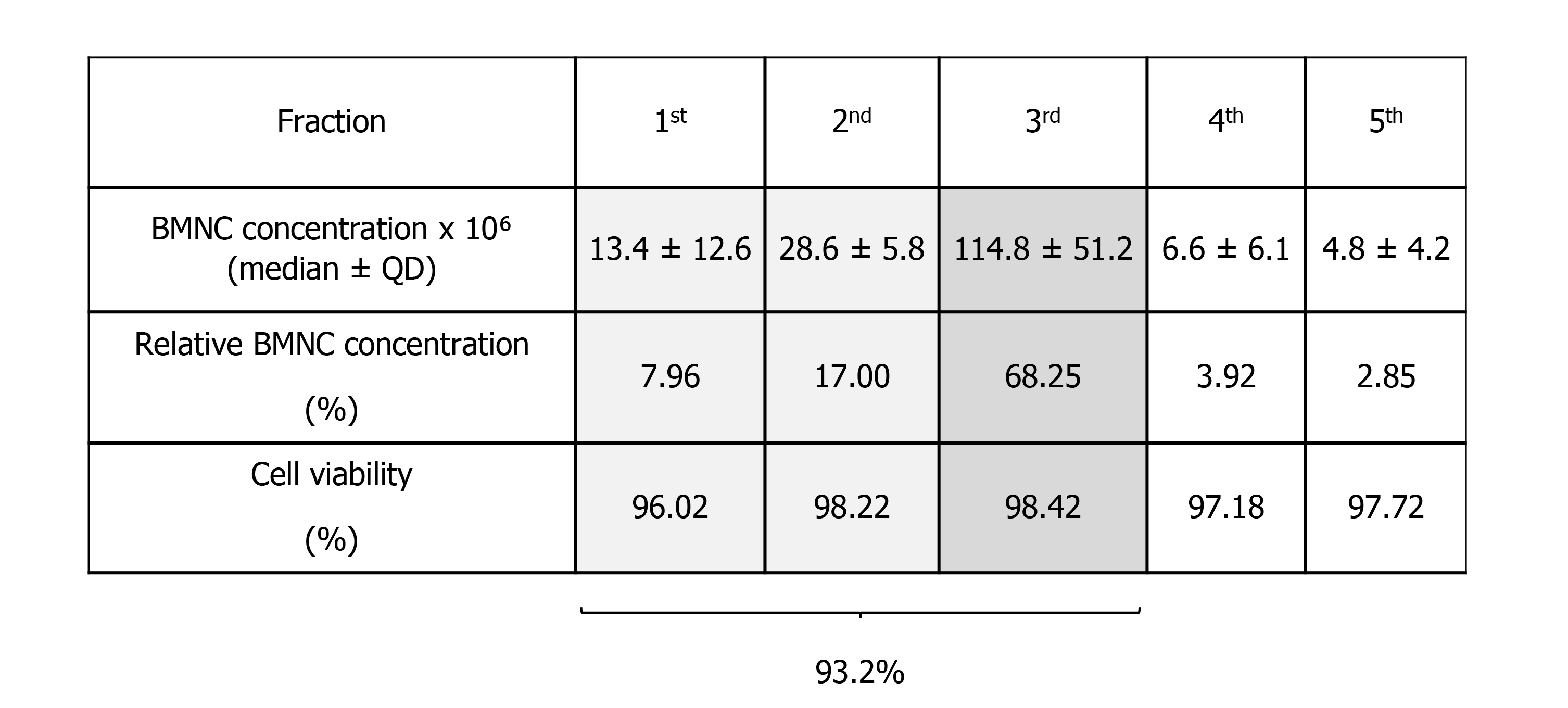
Figure 4 Mononuclear cell content in five consecutive bone marrow aspirates (5 mL each).
The first three fractions represent 93.2% of the total 25 mL (the five fractions together) emphasizing the need to obtain at least the first 15 mL of bone marrow aspirate. BMNC: Bone marrow mononuclear cells.
- Citation: Menarim BC, MacLeod JN, Dahlgren LA. Bone marrow mononuclear cells for joint therapy: The role of macrophages in inflammation resolution and tissue repair. World J Stem Cells 2021; 13(7): 825-840
- URL: https://www.wjgnet.com/1948-0210/full/v13/i7/825.htm
- DOI: https://dx.doi.org/10.4252/wjsc.v13.i7.825


