Published online Mar 15, 2003. doi: 10.3748/wjg.v9.i3.606
Revised: December 12, 2002
Accepted: December 22, 2002
Published online: March 15, 2003
AIM: To evaluate systematically our nine-year experience in treating right-sided diverticulitis of the colon, and to explore its clinical and radiological relationship.
METHODS: The clinical and radiological data of 40 patients with colonic diverticulitis treated in Mackay Memorial Hospital, Taipei, from 1993 through 2002 were reviewed retrospectively.
RESULTS: The average age of the patients with right-sided diverticulitis was 53.1 years, which was 11.6 years younger than that of the patients with left-sided diverticulitis. The preoperative diagnosis of appendicitis was made in 8 of 13 right-sided diverticulitis patients. Nine (69%) had right lower quadrant abdominal pain for more than 48 hours, and ten patients (77%) presented with fever. CT findings suggesting acute right-sided diverticulitis including thickening of the intestinal wall and pericolonic inflammation were present in five patients.
CONCLUSION: Right-sided diverticulitis is easily confused with acute appendicitis because it occurs at a somewhat younger age than that in left-sided diverticulitis. Barium enema and CT are helpful for the early diagnosis of right-sided diverticulitis. While clearly not required in the majority of patients with right lower quadrant abdominal pain, barium enema and CT may be helpful in making the decision with a clinical history or physical examinations atypical of acute appendicitis.
- Citation: Shyung LR, Lin SC, Shih SC, Kao CR, Chou SY. Decision making in right-sided diverticulitis. World J Gastroenterol 2003; 9(3): 606-608
- URL: https://www.wjgnet.com/1007-9327/full/v9/i3/606.htm
- DOI: https://dx.doi.org/10.3748/wjg.v9.i3.606
Diverticular disease was almost unknown in 1990, but has become the commonest affliction of the colon in Western countries[1] which was regarded as a deficient disease of Western civilization. The right-sided diverticulitis is more common than left-sided diverticulitis in Far-eastern countries. In a study of 105 patients in Taiwan, China, the incidence of right-sided diverticulosis was 60%[2]. The distinction between right-sided diverticulitis and acute appendicitis is often difficult at the time of presentation[3]. The condition is frequently misdiagnosed and has often been mistreated. During the past nine years, we have treated 13 cases of right-sided diverticulitis at the Mackay Memorial Medical Center, our experience in relation to the clinical and radiological manifestations was reviewed.
The pathological reports of medical records at the Mackay Memorial Medical Center were reviewed from January 1993 to June 2002. During this period, there were 40 patients with colonic diverticulitis treated at our institution, we retrospectively reviewed their presentation, diagnostic studies, management and pathology. Colonic diverticulitis was stratified into two groups according to the distribution of diverticula: (1) right-sided diverticulitis with diverticula in the cecum, ascending colon and proximal transverse colon; (2) left-sided diverticulitis with diverticula in the sigmoid and/or descending colon. Clinical details of these patients were shown in Table 1. Presenting symptoms and signs of right-sided diverticulitis patients were shown in Table 2.
| Right-sided diverticulitis | Left-sided diverticulitis | |
| Number | 13 | 27 |
| Male/Female | 10/3 | 20/7 |
| Age (yr) | ||
| Mean ± SD | 53.15 ± 9.86 | 64.74 ± 11.28 |
| Range | 40-70 | 44-83 |
| Median | 50 | 68 |
| Initial features | Cases number (%) |
| RLQ abdominal pain for > 2 days | 9(69) |
| Nausea/vomiting | 2(15) |
| Diarrhea | 2(15) |
| Leukocytosis | 9(69) |
| Fever | 10(77) |
| Anorexia | 1(8) |
Statistical comparison was performed using Student’s t test. All analyses were performed with the Stastical Package for the Social Science (SPSS) for windows (Version 10.0) software. The results were considered to be statistically significant at a value of P < 0.05.
The incidence of right-sided diverticulitis was 33% in our treated patients. There was no difference between right-sided diverticulitis patients and left-sided diverticulitis patients as regarding to male: female ratio. The age of patients with right-sided diverticulitis was younger than those with left-sided diverticulitis (P = 0.003, Table 1).
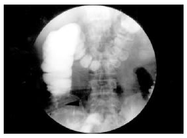
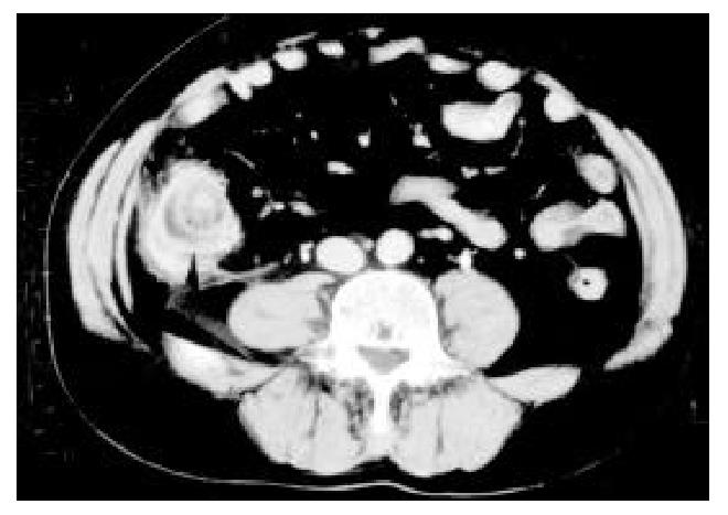
Details of their presenting symptoms and signs were shown in Table 2. The primary complaints among all patients were right lower quadrant abdominal pain for more than two days prior to admission. Nausea and/or vomiting were reported in two patients (15%) and diarrhea was present in two patients (15%), but only one of the 13 exhibited anorexia. Ten patients had a fever of more than 37 °C and all of these also had a leukocytosis. The preoperative diagnosis was appendicitis in 8 of the 13 patients in our series. Four patients had performed preoperative barium enemas. In one patient a 4 cm × 4.5 cm diverticulum of the proximal transverse colon was identified. In the other three, the barium enema was interpreted as showing a right-sided colonic mass (Figure 1). Six patients had CT scans, of which five correctly diagnosed diverticulitis of the cecum. Diverticulitis of the right colon was correctly diagnosed preoperatively in one patient in whom marked wall thickening of the cecum with classic target appearance was demonstrated (Figure 2). Diverticula was found in one patient. There was no evidence of contrast extravasation in any of the six patients.
The preoperative distinction between right-sided diverticulitis and appendicitis is extremely difficult to discern based on clinical presentation alone[4]. In our study, a misdiagnosis of appendicitis was made in 8 of the 13 patients preoperatively which is common in all other reported series[3-5]. The majority of patients will still undergo laparotomy. The surgeon must make a diagnosis based on operative findings. Hidden diverticula pose a diagnostic dilemma since at laparotomy it may be difficult to distinguish the inflamed mass from Crohn’s disease, malignant lesions of the right colon, a perforated foreign body or even tuberculosis[3].
Patients with right-sided diverticulitis tend to be younger than those with left-sided diverticulitis[5]. We compared the age distribution of right-sided diverticulitis and left-sided diverticulitis patients in this study (Table 1).
Clinically, in contrast to appendicitis, relative long history of right lower quadrant abdominal pain, relative lack of systemic toxic signs and low frequency of nausea/vomiting may be helpful in correctly diagnosing right-sided diverticulitis[2]. The symptoms of right-sided diverticulitis usually begin and remain localized in the right lower quadrant, rather than originating in the epigastrium[6]. Appendicitis patients typically experience the classic migration of pain to the right lower quadrant at later stage which caused by the stimulation of the visceral afferent nerve fibers that enter the spinal cord at thoracic levels T8 through T10. Nine of our patients presented more than 48 hours after the onset of symptoms (69%). Eleven of our patients (85%) had neither nausea, vomiting, nor diarrhea. This absence of vomiting has been noted by others[7-9].
About one third of patients in this series were right-sided diverticulitis, which differed from other reports in Far-eastern countries[10-12]. This bia was due to our review was obtained from pathological database of medical records from our hospital, in which some uncomplicated right-sided diverticulitis cases were excluded. Right-sided diverticulitis tends to have a more benign course than that which occurs on the left[13]. So this differential probably carries little significance, despite our own findings. Diverticular disease is considered as a fiber deficient disease in Western Countries[1], so duty of the profession is to point the way of prevention of white flour, both brown and white sugar, confectionery, and foods or drinks which contain unnaturally concentrated carbohydrates. But some Eastern racial groups have a higher incidence of right-sided diverticulitis despite a high fiber intake. These findings were coincident to a study from China where about 62% of right-sided diverticular disease despite a good fiber intake[14].
Contrast enema studies are the most accurate way to find out the colonic diverticula[15]. However, because of the risks of extravasation of barium from the perforation in the patients with acute right-sided diverticulitis, barium enema examination should be generally be avoided in patients with suspected acute right-sided diverticulitis and localized peritoneal signs. Criterias for the diagnosis of right-sided diverticulitis include extravasation of barium, narrowed lumen or thickened mucosa, and mass effect[15]. In our series, barium enema studies were done in four patients who presented a cecal mass in three patients.
In contrast to the barium enema, CT scan demonstrates both the intraluminal and extracolonic manifestations of acute right-sided diverticulitis[16]. Criterias of CT scan for the diagnosis of right-sided diverticulitis include colonic wall thickening, pericolonic fat infiltration (streaky fat), pericolonic or distant abscesses, and extraluminal air. In this series, CT scans were obtained preoperatively in six patients with right-sided diverticulitis. CT findings suggesting right-sided diverticulitis were present in five patients. A recent study found that pericolonic lymph nodes adjacent to the focal area of colonic thickening are more commonly seen in patients with colonic cancer. Pericolonic inflammatory changes are more commonly seen in right-sided diverticulitis[17]. CT may be helpful for the evaluation of patients with atypical symptoms of acute appendicitis or those who have undergone an appendectomy. Right-sided diverticulitis occurs with greater frequency in Asians. This condition is easily confused with acute appendicitis, since it occurs at a somewhat younger age than those with left-sided diverticulitis. If dignosed preoperatively, uncomplicated right-sided diverticulitis can be managed conservatively with antibiotic therapy.
Edited by Xu XQ
| 1. | Painter NS, Burkitt DP. Diverticular disease of the colon: a deficiency disease of Western civilization. Br Med J. 1971;2:450-454. [RCA] [PubMed] [DOI] [Full Text] [Cited by in RCA: 1] [Reference Citation Analysis (0)] |
| 2. | Chiu JH, Lin JT, Lin JK, Leu SY, Liang CL, Wang FM. Diverticu-lar disease of the colon. J Surg Asso. 1987;20:102-108. |
| 3. | Gouge TH, Coppa GF, Eng K, Ranson JH, Localio SA. Management of diverticulitis of the ascending colon. 10 years' experience. Am J Surg. 1983;145:387-391. [PubMed] |
| 4. | Markham NI, Li AK. Diverticulitis of the right colon--experience from Hong Kong. Gut. 1992;33:547-549. [RCA] [PubMed] [DOI] [Full Text] [Cited by in Crossref: 79] [Cited by in RCA: 69] [Article Influence: 2.1] [Reference Citation Analysis (0)] |
| 5. | Fischer MG, Farkas AM. Diverticulitis of the cecum and ascending colon. Dis Colon Rectum. 1984;27:454-458. [RCA] [PubMed] [DOI] [Full Text] [Cited by in Crossref: 49] [Cited by in RCA: 46] [Article Influence: 1.1] [Reference Citation Analysis (0)] |
| 6. | Birnbaum BA, Wilson SR. Appendicitis at the millennium. Radiology. 2000;215:337-348. [RCA] [PubMed] [DOI] [Full Text] [Cited by in Crossref: 401] [Cited by in RCA: 343] [Article Influence: 13.7] [Reference Citation Analysis (0)] |
| 7. | Arrington P, Judd CS. Cecal diverticulitis. Am J Surg. 1981;142:56-59. [RCA] [PubMed] [DOI] [Full Text] [Cited by in Crossref: 29] [Cited by in RCA: 28] [Article Influence: 0.6] [Reference Citation Analysis (0)] |
| 8. | Asch MJ, Markowitz AM. Cecal diverticulitis: report of 16 cases and a review of the literature. Surgery. 1969;65:906-910. [PubMed] |
| 9. | Schuler JG, Bayley J. Diverticulitis of the cecum. Surg Gynecol Obstet. 1983;156:743-748. [PubMed] |
| 10. | Wang CH, Chou LC. The incidence of the diverticular diseases of the colon in T.S.G.H., Taiwan, China. J Surg Asso. 1979;12:260-266. |
| 11. | Sugihara K, Muto T, Morioka Y, Asano A, Yamamoto T. Diverticular disease of the colon in Japan. A review of 615 cases. Dis Colon Rectum. 1984;27:531-537. [RCA] [PubMed] [DOI] [Full Text] [Cited by in Crossref: 130] [Cited by in RCA: 114] [Article Influence: 2.8] [Reference Citation Analysis (0)] |
| 12. | Vajrabukka T, Saksornchai K, Jimakorn P. Diverticular disease of the colon in a far-eastern community. Dis Colon Rectum. 1980;23:151-154. [RCA] [PubMed] [DOI] [Full Text] [Cited by in Crossref: 40] [Cited by in RCA: 37] [Article Influence: 0.8] [Reference Citation Analysis (0)] |
| 13. | Ferzoco LB, Raptopoulos V, Silen W. Acute diverticulitis. N Engl J Med. 1998;338:1521-1526. [RCA] [PubMed] [DOI] [Full Text] [Cited by in Crossref: 251] [Cited by in RCA: 200] [Article Influence: 7.4] [Reference Citation Analysis (0)] |
| 14. | Pan GZ, Liu TH, Chen MZ, Chang HC. Diverticular disease of colon in China. A 60-year retrospective study. Chin Med J (Engl). 1984;97:391-394. [PubMed] |
| 15. | Beranbaum SL, Zausner J, Lane B. Diverticular disease of the right colon. Am J Roentgenol Radium Ther Nucl Med. 1972;115:334-348. [RCA] [PubMed] [DOI] [Full Text] [Cited by in Crossref: 25] [Cited by in RCA: 21] [Article Influence: 0.4] [Reference Citation Analysis (0)] |
| 16. | Crist DW, Fishman EK, Scatarige JC, Cameron JL. Acute diverticulitis of the cecum and ascending colon diagnosed by computed tomography. Surg Gynecol Obstet. 1988;166:99-102. [PubMed] |
| 17. | Macari M, Balthazar EJ. CT of bowel wall thickening: significance and pitfalls of interpretation. AJR Am J Roentgenol. 2001;176:1105-1116. [PubMed] [DOI] [Full Text] |


