Published online Oct 15, 2000. doi: 10.3748/wjg.v6.i5.676
Revised: June 16, 2000
Accepted: June 23, 2000
Published online: October 15, 2000
AIM: To investigate the morphological and ultrastructural changes in the human gastric carcinoma cell line BGC-823 after being treated with tachyplesin.
METHODS: Tachyplesin was isolated from acid extracts of Chinese horseshoe crab (Tachypleus tridentatus) hemocytes. BGC-823 cells and the cells treated with 2.0 mg/L tachyplesin were examined respectively under light microscope, scanning and transmission electron microscope.
RESULTS: BGC-823 cells had undergone the restorational alteration in morphology and ultrastructure after tachyplesin treatment. The changes were as follows: the shape of cells was unanimous, the volume enlarged and cells turned to be flat and spread, the nucleo-cytoplasmic ratio lessened and nuclear shape became rather regular, the number of nucleolus reduced and its volume lessened, heter-chromatin decreased while euchromatin increased in nucleus. In the cytoplasm, mitochondria grew in number with consistent structure relatively, Golgi complex turned to be typical and well-developed, rough endoplasmic reticulum increased and polyribosome decreased. The microvilli at cellular surface were rare and the filopodia reduced while lamellipodia increased at the cell edge.
CONCLUSION: Tachyplesin could alter the malignant morphological and ultrastructural characteristics of human gastric carcinoma cells effectively and have a certain inducing differen-tiation effect on human gastric carcinoma cells.
- Citation: Li QF, Ou-Yang GL, Li CY, Hong SG. Effects of tachyplesin on the morphology and ultrastructure of human gastric carcinoma cell line BGC-823. World J Gastroenterol 2000; 6(5): 676-680
- URL: https://www.wjgnet.com/1007-9327/full/v6/i5/676.htm
- DOI: https://dx.doi.org/10.3748/wjg.v6.i5.676
Horseshoe crab, a kind of marine animals, is a “live fossil” which has an unique status in evolution. It has many bioactive substances with special functions due to its primitive character. In recent years, many bioactive substances, including clotting factors, protease inhibitors, antibacterial substances, lectins and others, have been found in the hemocytes and hemolymph plasma of horseshoe crab[1]. To seek the low molecular weight antitumor substances which can intervene in cellular signal transduction and regulate cell proliferation and differentiation[2], we have isolated and purified a small polypeptide - tachyplesin from the blue blood of Chinese horseshoe crab (Tachypleus tridentatus) and appraised its antitumor activities[3]. Then we have investigated the biological effects of tachypelsin on tumor cells systematically with the model system of human gastric carcinoma cell line BGC-823. This paper deals with the effects of tachyplesin on the morphology and ultrastructure of human gastric carcinoma cells by light microscopy and scanning and transmission electron microscopy.
Tachyplesin was isolated from acid extracts of hemocyte debris of Chinese horseshoe crab as described by Nakamura[4] with minor modification.
BGC-823 cells were cultured in RPMI-1640 supplemented with 20% heat-inactivat ed fetal calf serum, 100 units/mL penicillin, 100 mg/L streptomycin and 50 mg/L kanamycin at 37 °C, 5% CO2 in air. Then the cells were treated with the culture medium containing 2.0 mg/L tachyplesin after being seeded for 24 h.
BGC-823 cells and the cells treated with 2.0 mg/L tachyplesin were seeded in little penicillin bottles with cover slip strip, and grown in the normal culture medium or in the culture medium containing 2.0 mg/L tachyplesin at 37 °C in 5% CO2 atmosphere for 72 h respectively. The cells at cover slip strips were rinsed with D-Hank’s solution twice at 37 °C, fixed overnight in Bouin-Hollande fixative, stained with Hematoxylin-Eosin, and observed under light microscope.
BGC-823 cells culture and tachyplesin treatment were performed as the procedures for light microscopy. The cells at cover slip strips were rinsed with D-Hank’s solution twice at 37 °C, fixed in 2.5% glutaraldehyde for 2 h and in 1% osmian tetroxide for 1 h, dehydrated in ethanol, dried through the CO2 critical point, gilded in vacuum and observed under the HITIACHS-520 scanning electron microscope.
BGC-823 cells and the cells treated with 2.0 mg/L tachyplesin were rinsed with D-Hank’s solution twice, shaved into centrifuge tube with plastic scraper, followed by centrifugation at 2000 rpm for 15 min, and removed the supernatant. The precipitate was prefixed in 2.5% glutaraldehy de for 2 h and postfixed in 1% osmian tetroxide for 2 h, dehydrated in ethanol series, embedded in epoxy resin 618, stained with lead citrate and uranyl acetate, and observed under the JEM-100CXII transmission electron microscope.
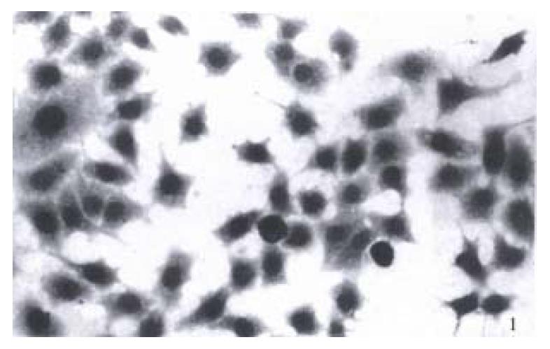
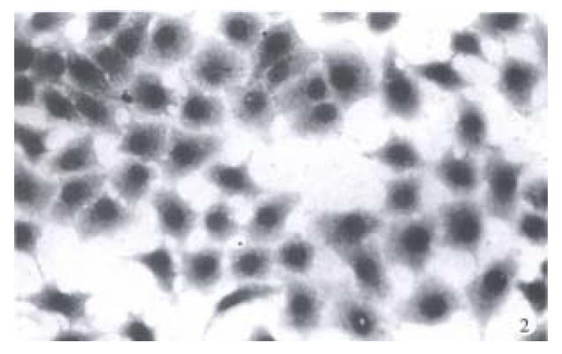
There were various forms in BGC-823 cells, such as epithelioid, round, shuttle-like and irregular shapes, as well as cancerous giant cells and soon. The volume of BGC-823 cells was small relatively, nucleus was large and irregular with several nucleoli in it, and cytoplasm was few (Figure 1). After being treated with 2.0 mg/L tachyplesin, BGC-823 cells had undergone a significant morphological change and appeared as normal differentiated epithelial cells. The cells tended to be spread and flat apparently, their volume enlarged, cytoplasm was abundant, nucleus became relatively smaller with its shape rather regular, and the number of nucleoli was fewer. This feature was different from that of BGC-823 cells remarkably (Figure 2).
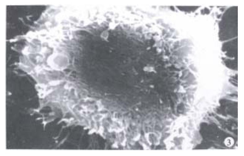
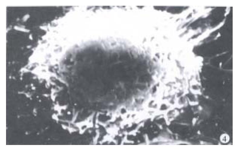
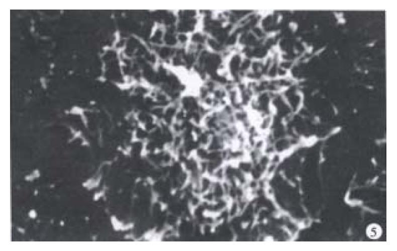
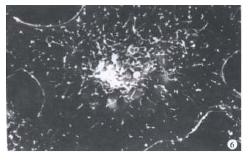
Under the scanning electron microscope, BGC-823 cells presented on spherical, shuttle-like and flat and spread shape. There were abundant microvilli at the surface of each type of cells. Microvilli were distributed densely at the surface of spherical cells while densely at the center and scattered at the edge of spread cells. Filopodia were abundant and arranged radiately, especially at the edge of spherical cells while there were relatively few filopodia and a little lamellipodia at the edge of spread cells (Figure 3, Figure 4, Figure 5). In contrast, the BGC-823 cells, after being treated with 2.0 mg/L tachyplesin, also appeared in spherical, shuttled-like and flat and spread shapes, while the spread cells grew in number and spherical cells reduced. The cellular surface appeared rather smooth with a few small bubble and ruffled structure. The filopodia at cellular edge lessened and lamellipodia increased, especially large lamellipodia could be seen at the edge of flat and in spread cells. This surface feature was obviously different from that of BGC-823 cell (Figure 4, Figure 6).
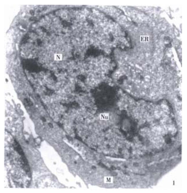
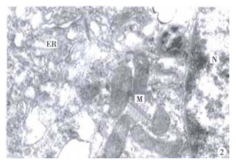
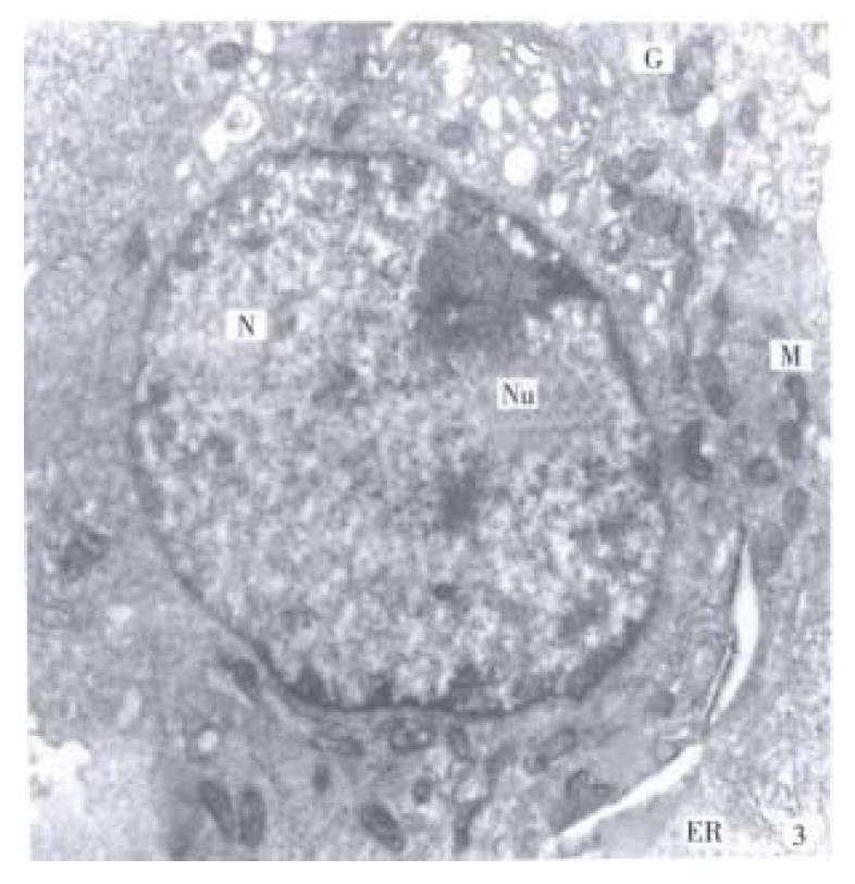
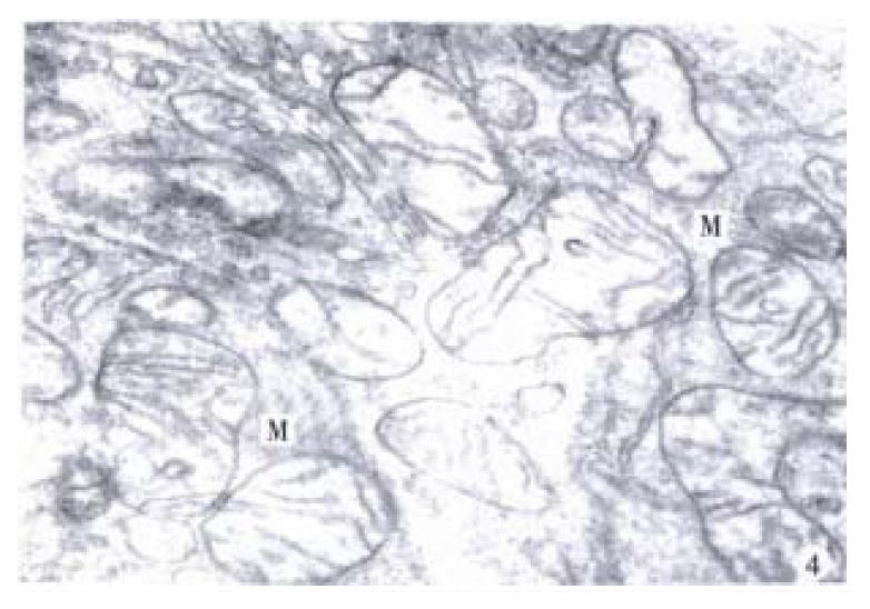
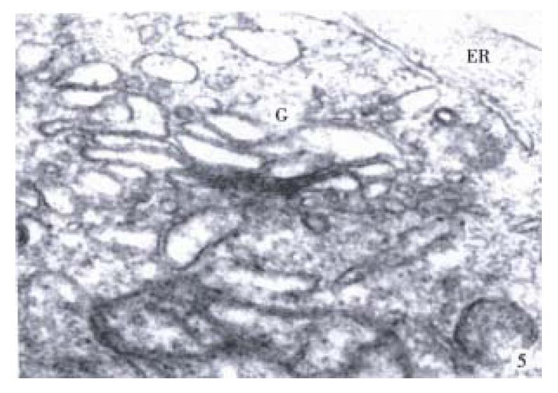
It was revealed by transmission electron microscope that the necleo-cytoplasmic ratio of BGC-823 cells was relatively large, nucleus was irregular, many heterochromatins in the nucleus and nucleolus were large, varied in shape and with some nucleolus vacuoles. Meanwhile, rough endoplasmic reticulum was not well-developed, Golgi vesicle was few, arranged irregularly and Golgi cisterna swelled obviously, mitochondria were irregular and cristae within mitochondria arranged irregularly, polyribosome were abundant while free ribosome were few (Figure 7, Figure 8). However, after 2.0 mg/L tachyplesin treatment, the ultrastructure of BGC-823 cells had also undergone a significant change. The necleo-cytoplasmic ratio lessened, the nuclear shape became regular, most being round or oval, heterochromatin in nucleus decreased while euchromatin increased, the volume of nucleolus lessened, rough endoplasmic reticulum increased obviously, Golgi apparatus was well-developed and Golgi vesicle increased and arranged regularly, most of mitochondria were oval, and their cristae grew in number and arranged regularly, polyribosome reduced while free ribosome increased (Figure 9, Figure 10, Figure 11). BGC-823 cells showed some ultrastructural characteristics of their normal relevant cells after being treated with tachyplesin.
There was significant difference in morphology and ultrastructure between tumor cells and their relevant normal cells. Tumor cells usually display some malignant morphological and ultrastructural characteristics such as that the nucleo-cytoplasmic ratio was relatively large, nucleus was large and malformed with several nucleoi in it, cell organelle were not well-developed and microvilli were abundant[5]. Therefore, to observe and identify the changes of morphology and ultrastructure in tumor cells is important in determining the effects of exotic substances especially differentiational inducers on tumor cells[6]. A series of research in which differentiation of gastric carcinoma, leukemia, hepatoma and lung carcinoma cells were induced by chemical inducers, have made it clear that the morphology and ultrastructure of carcinoma cells after induced treatment engendered a restorative alteration similar to those of relevant normal cells[7-11].
BGC-823 cell is a poorly-differentiated human gastric adenocarcinoma cell line with fast-proliferation and high-malignant characteristics. Our results showed that BGC-823 cells had the typical malignant phenotypical characteristics of morphology and ultrastructure in tumor cells. However, in the BGC-823 cells after treatment with 2.0 mg/L tachyplesin, it displayed the changes as follows: the cells were unanimous and tended to be spread and became flat apparently, the volume of cells enlarged, epithelioid-like cells grew in number, the necleo-cytoplasmic ratio declined, the shape of nucleus became relatively regular, the volume and number of nucleolus decreased, heterochromatin decreased while euchromatin increased, mitochondria and their cristae increased, Golgi complex was well-developed and typical, rough endoplasmic reticulum increased, polyribosome decreased while free ribosome increased, microvilli and filopodia decreased while lamellipodia increased. These changes were different from the morphology and ultrastructure of BGC-823 cells and similar to those of their relevant normal cells. It in dicated that tachyplesin could alter the malignant phenotypical characteristics of morphology and ultrastructure in gastric carcinoma cells and make them take on the morphological and ultrastructural characteristics similar to those of their normal cells.
The action of tachyplesin is identical with those of chemical inducers on gastric carcinoma cells. It was reported that the human gastric carcinoma cell line SGC-7901 treated with sodium butyrate[12], hexamethylemene bisacetamide (HMBA)[13] and all-trans retinoic acid[14,15]. The shape of cell was rather regular and consistant, the nuclear shape became round and nucleolus shrank, the nucleo-cytoplasmic ratio lessened and hetrochromatin reduced, and the organelle were well-developed, and microvilli at cell surface decrease. These changes were unanimous with the changes of tachyplesin on gastric carcinoma cells in this paper. It further demonstrates that tachyplesin has the identical action with differential inducers of cancer cells, and it has a certain inducing differential effect on human gastric carcinoma cells. In this connection, it has a momentous significance for application of tachyplesin in tumor treatment and in the antitumor research of marine bioactive substance to study the acting mechanism of tachyplesin on tumor cells further.
Edited by You DY Verified by Ma JY
| 1. | Iwanaga S, Kawabata S, Muta T. New types of clotting factors and defense molecules found in horseshoe crab hemolymph: their structures and functions. J Biochem. 1998;123:1-15. [RCA] [PubMed] [DOI] [Full Text] [Cited by in Crossref: 226] [Cited by in RCA: 198] [Article Influence: 7.3] [Reference Citation Analysis (0)] |
| 2. | Johnson TC. Negative regulators of cell proliferation. Pharmacol Ther. 1994;62:247-265. [RCA] [PubMed] [DOI] [Full Text] [Cited by in Crossref: 19] [Cited by in RCA: 22] [Article Influence: 0.7] [Reference Citation Analysis (0)] |
| 3. | Hong SG, Chen F, Li QF, Hu YC, Ye J, Ouyang GL, Li CY, Li XQ, Qiao YH, Chen F. Study on antitumor activity of tachyplesin-I against human promyelocytic leukemia cell line HL 60. Xiamen Daxue Xuebao (Ziran Kexue Ban). 1999;38:448-451. |
| 4. | Nakamura T, Furunaka H, Miyata T, Tokunaga F, Muta T, Iwanaga S, Niwa M, Takao T, Shimonishi Y. Tachyplesin, a class of antimicrobial peptide from the hemocytes of the horseshoe crab (Tachypleus tridentatus). Isolation and chemical structure. J Biol Chem. 1988;263:16709-16713. [PubMed] |
| 5. | Symington T, Carter RL. Scientific foundations of oncology. Beijing: Kexue Chubanshe 1984; 11-18. |
| 6. | Jing YK. The research advance in induing differentiation of cancer cells and the differentiational inducers. Yaoxue Jinzhan. 1992;16:6-13. |
| 7. | Li XG, Xie JY, Lu YY. Suppressive action of garlic oil on growth and differentiation of human gastric cancer cell line BGC 823. World J Gastroentero. 1998;4:13. |
| 8. | Li QF. Effect of retinoic acid on the changes of nuclear matrix in termediate filament system in gastric carcinoma cells. World J Gastroenterol. 1999;5:417-420. [PubMed] |
| 9. | Ryves WJ, Dimitrijevic S, Gordge PC, Evans FJ. HL-60 cell differentiation induced by phorbol- and 12-deoxyphorbol-esters. Carcinogenesis. 1994;15:2501-2506. [RCA] [PubMed] [DOI] [Full Text] [Cited by in Crossref: 8] [Cited by in RCA: 9] [Article Influence: 0.3] [Reference Citation Analysis (0)] |
| 10. | Vesey DA, Cunningham JM, Selden AC, Woodman AC, Hodgson HJ. Dimethyl sulphoxide induces a reduced growth rate, altered cell morphology and increased epidermal growth factor binding in Hep G2 cells. Biochem J. 1991;277:773-777. |
| 11. | Khan MZ, Freshney RI, McNicol AM, Murray AM. Induction of phenotypic changes in SCLC cell lines in vitro by hexamethylene bisacetamide, sodium butyrate, and cyclic AMP. Annals Oncology. 1993;4:499-507. |
| 12. | Lü GZ, Gao Y, Huang YC, Lin ZX, Wang KR. [The biological effect of sodium butyrate (NaBT) on SGC-7901 cells]. Shiyan Shengwu Xuebao. 1989;22:169-175. [PubMed] |
| 13. | Xu SW, Zhao HY, Ren LQ, Pei ZL. Electron microscope observation on the induced differentiation of human gastric adenocarcinoma cell line SGC 7901 by HMBA. Zhongliu. 1989;9:260-261. |
| 14. | Xia F, Wang DK, Liu BH, Feng SZ, Chen L, Li YB. Effects of differenti ation inducers in combination with cytotoxic agents on human gastric carcinoma cell line SGC 7901. Dier Junyi Daxue Xuebao. 1998;20:220-223. |
| 15. | Chen Y, Xu CF. All transretinoic acid induced differentiation in human gastric carcinoma cell line SGC 7901. Xin Xiaohuabingxue Zazhi. 1997;5:491-492. |


