Review Open Access
Copyright ©The Author(s) 2000. Published by Baishideng Publishing Group Inc. All rights reserved.
World J Gastroenterol. Aug 15, 2000; 6(4): 465-469
Published online Aug 15, 2000. doi: 10.3748/wjg.v6.i4.465
Molecular biology of liver disorders: the hepatitis C virus and molecular targets for drug development
Howard J. Worman, Feng Lin, Departments of Medicine and of Anatomy and Cell Biology, College of Physi cians and Surgeons, Columbia University, 630 West 168th Street, New York, NY 100 32, USA
Howard J. Worman is Associate Professor of Medicine and Anatomy and Cell Biology at Columbia University’s College of Physicians and Surgeons and Director of the Division of Digestive and Liver Diseases of the Medical Service at the New York-Presbyterian Hospital Columbia-Presbyterian Campus. He has over 70 scientific papers as well as publications on liver diseases for the lay audience.
Author contributions: All authors contributed equally to the work.
Correspondence to: Dr. Howard J. Worman, Depar tment of Medicine, College of Physi cians and Surgeons, Columbia University, 630 West 168th Street, New York, NY 10032, USA. hjw14@columbia.edu
Telephone: +1-212-3058156 Fax: +1-212-3056443
Received: May 25, 2000
Revised: June 12, 1999
Accepted: June 15, 2000
Published online: August 15, 2000

Abstract
Key Words: hepatitis C, molecular biology, viral hepatitis, anti-viral agents


INTRODUCTION

Molecular biology has made a tremendous impact on the diagnosis and treatment of liver diseases[1,2]. In particular, advances in molecular biology made possible the discovery of the virus that causes hepatitis C. In this review, we use hepatitis C as an example of the impact that molecular biology has made in t he area of liver disorders. We emphasize how our growing understanding of the he patitis C virus (HCV) has lead to the identification of targets for development of new treatments.

THE HEPATITS C VIRUS (HCV)
Basic molecular virology of HCV

Investigators at Chiron Corporation were the first to discover HCV and reported this in a landmark paper published in 1989[3]. The virus was identified by antibody screening of cDNA expression libraries made from DNA and RNA from the plasma of chimpanzees. These chimpanzees were inoculated with serum from human s with what was then called post-transfusion “non-A, non-B” hepatitis. The DNA expression library was screened with antibodies from sera of other patients with “non-A, non-B” hepatitis. This led to the isolation of clones that we re derived from portions of the viral genome and encoded fragments of viral poly peptides. Treatment with RNase and DNase showed that HCV was a positive-stranded RNA virus[3]. In an accompanying paper, the investigators who discover ed HCV and their collaborators showed that the vast majority of individuals with chronic “non-A, non-B hepatitis” had antibodies against the newly identifi ed viral polypeptides[4].

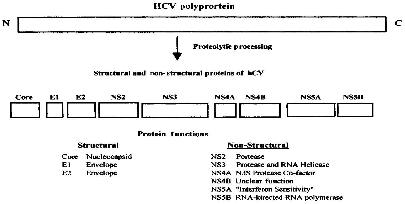
After the discovery of HCV, its entire genome was cloned and sequenced in several laboratories[5-8]. HCV is a member of the Flavivaridae family. Once HCV infects cells, the positive, single stranded RNA genome is translated into a polyprotein of 3010 to 3033 amino acids, depending upon the strain (Figur e 1). The viral RNA is not capped and translation occurs via an internal ribosome entry site (IRES) at the 5’ end of the viral RNA[9,10]. The mechanis m of translation of uncapped viral RNA therefore differs from that used by virtu ally all cellular mRNAs which are capped at their 5’ ends.

Both host cell and viral proteases cleave the HCV polyprotein into severa l smaller polypeptides (Figure 1). The major structural proteins are core prote in and two envelope proteins called E1 and E2. Core protein forms the nucleocap sid of the mature virion and E1 and E2 are present in the viral envelope. A small polypeptide called P7 is also generated as a result of cleavage at the E2-NS 2 junction but its function is not clear. Four major non-structural proteins called NS2, NS3, NS4, and NS5 are generated, two of which, NS4 and NS5, are furth er processed into smaller polypeptides called NS4A, NS4B, NS5A, and NS5B. The non-structural proteins have enzymatic functions that are critical for viral replica tion in cells, such as RNA helicase (NS3), protease (NS2, NS3-NS4A complex) and RNA polymerase (NS5B) activities. NS5A has been implicated in determining sensi tivity to interferon alpha.

Figure 1
Figure 1 HCV proteins and their functions. The positive-stranded RNA of about 10000 nuc leotides is translated into a polyprotein of approximately 3000 amino acids. The polyprotein is proteolytically cleaved into several smaller proteins. Core, E1, and E2 are structural polypeptides. Core protein is the virus nucleocapsid and E 1 and E2 are viral envelope proteins. A small polypeptide known as P7 (not shown ) is also produced by additional cleavage between E2 and NS2. The major non-str uctural proteins are NS2, NS3, NS4, and NS5. NS4 is further processed into NS4A a nd NS4B and NS5 into NS5A and NS5B. NS2 and part of NS3 are proteases that proce ss the viral polyprotein. NS3 also has RNA-helicase activity. NS4A is a cofacto r for the NS3 protease and NS5B is an RNA-dependent, RNA polymerase. The functi ons of NS4B and NS5A are less well understood but NS5A is thought to play a role in determining sensitivity to interferon.
HCV replication and interactions with host cells

Little is known about the fundamental aspects of HCV replication, primarily beca use a robust cell culture has not been established. Although viral proteins and RNA components involved in critical steps in HCV replication are known, very li ttle is understood about the mechanistic details or the role of accessory host cell factors. Some of the basic steps in HCV replication that occur in infected cells are outlined here.

After infection of cells, HCV RNA must be translated into protein. HCV RN A translation is initiated by internal ribosome binding, not by 5’-end depend ent mechanisms[9,10]. Internal initiation is specified by an IRES ele me nt. Such elements were first discovered in the genomes of picornaviruses[11]. The IRES is believed to require the set of canonical translation initiation factors in order to function. In addition, IRES function is also thought to be dependent on other cell proteins. However, no single cell protein has been shown to be dispensable for the function of all IRESes.

HCV RNA must be unwound for efficient protein synthesis to occur. This process is catalyzed by a RNA helicase that is part of the viral NS3 protein. The three -dimensional structure of the HCV NS3 helicase domain has been determined and details about its function are emerging[12-14]. At the present time, it is not known if host cell co-factors are necessary for optimal functioning of the NS3 helicase. Cellular RNA helicases have also been shown to bind to the HCV core protein[15-17], however, it is not known if they also play a role in unwinding viral RNA.

After its synthesis, the HCV polyprotein is processed into the structural and no nstructural proteins. Proteolytic cleavages between structural polypeptides are catalyzed by signal peptidase in the endoplasmic reticulum. Two virally encode d proteases, NS2 and NS3, catalyze the other cleavages of the HCV polyprotein. The NS3 protease contains a trypsin-like fold and a zinc-binding site and is c omplexed with the viral protein NS4A[18,19].

HCV RNA must be replicated to produce more virions. The viral protein NS5B is an RNA-dependent RNA polymerase. NS5B bears some similarity and motif organizati on to poliovirus polymerase and human immunodeficiency virus 1 (HIV-1) reverse transcriptase but adopts a unique shape due to extensive interactions between th e fingers and thumb polymerase subdomains that encircle its active site[20]. The precise mechanism of action of the HCV NS5B polymerase is not known. C ellular or viral protein or RNA binding partners that function as subunits or in itiation factors may be necessary for optimal activity.

The replication rate of HCV in human hosts is estimated to be extremely high. It appears that the estimated half-life of a viral particle is 2.7 h with pr oduction and clearance of about one trillion viral particles a day[21] . This rate of virion production is approximately 1000 times greater than that estimated for HIV-1. Factors responsible for the high rate of HCV replication are not entirely understood. This rapid rate of replication can explain the develo pment of mutant strains or quasispecies that occur after HCV infection. It may a lso make development of an effective vaccine difficult.

DRUG TARGETS FOR THE TREATMENT OF HCV INFECTION
“Non-specific” anti-viral agents for HCV infection

The currently available drugs for the treatment of hepatitis C are anti-viral a gents not specifically directed against HCV. The United States Food and Drug Adm inistration (FDA) has approved several preparations of recombinant interferon al pha for the treatment of chronic hepatitis C. Interferon alpha is a suboptimal t reatment in that only about 20% or less of patients who complete a one year cour se of treatment respond successfully as determined by the inability to detect HCV in serum 6 mo after the drug is stopped[22]. Numerous adverse even ts are also associated with interferon alpha, most notably flu-like symptoms, neutropenia, thrombocytopenia, and depression. Interferon alpha must be administe red by injection 3 times a week. Newer preparations of interferon alpha-2b complexed with polyethylene glycol have been developed[23]. These so-cal led “PEG-ylated” interferon alphas are released more slowly and evenly into the bloodstream and need only be administered by injection once a week. “PEG -ylated” interferon alphas will likely be approved for the treatment of chron ic hepatitis C in the United States in the year 2000 or 2001.

The combination of interferon alpha-2b plusing oral ribavirin is approved in many countries for the treatment of chronic hepatitis C. Combination treatment for 6 mo leads to no detectable virus in serum 6 mo after stopping thera py in approximately 40% of subjects[24-26]. The major adverse event as sociated with ribavirin is hemolytic anemia, which in rare cases can be life thr eatening. VX-497 is a compound in development that inhibits inosine monophospha te dehydrogenase and may have antiviral affects similar to those of ribavirin[27]. VX-497 is being studied in combination with interferon alpha to e stablish if it is as effective as ribavirin with a similar or preferable adverse events profile.

Other cytokines have also been tested in the treatment of HCV infection. A recent report of a pilot study suggests that interleukin-10 may slow the deve lopment of liver fibrosis in subjects with chronic hepatitis C[28]. Inte rleukin-10, however, was not shown to have anti-viral activity against HCV.

Agents directed against HCV non-structural proteins

The next generation of drugs for the treatment of hepatitis C will likely be dir ected against non-structural HCV proteins with known enzymatic activities. Thre e major targets are the NS3 protease, NS3 helicase, and NS5B RNA-directed RNA polymerase. The fact that these proteins have enzymatic activities that can be mea sured in vitro make them amenable to high-throughput screening techniques f avored by pharmaceutical chemists. This obviates the need to grow HCV in cell cultures or in small animals, tasks that have eluded investigators.

The three-dimensional structure of the HCV NS3 protease domain has been determi ned by X-ray crystallography[18,19]. In addition, the structure of the NS3 protease domain complexed with an inhibitor has recently been established[29]. Armed with this knowledge, chemists can use rationale drug design to synthesis compounds to inhibit protease activity. Rational drug design can be c ombined with combinatorial chemistry in which a library of thousands or more str ucturally similar molecules is tested against the target. By combining rational drug design and combinatorial chemistry with high throughput screening techniques that measure enzymatic activity, N3S protease inhibitors can be identified, fu rther developed and ultimately tested in infected chimpanzees and humans.

Similar methods can be used to identify inhibitors of the N3S helicase domain and NS5B RNA dependent RNA polymerase. The three-dimensional structures of these proteins are also known[12-14]. Although human cells have RNA helicas es, their mechanism of action is probably different than RNA helicases of viruses[30]. Animal cells do not have RNA-dependent RNA polymerases, making N S5B an attractive target for an anti-viral agent.

Agents directed against HCV RNA

HCV RNA differs from cellular mRNA. First, it has a unique ribonucleotide seque nce. Second, as outlined above, HCV RNA is uncapped and translation is initiated via an IRES. Third, the viral RNA must be efficiently packaged into the matur e virions. These features make the HCV RNA a potential target for anti-viral drugs.

Ribozymes are catalytic RNA molecules that can be designed to cleave speci fic RNA sequences. Ribozymes therefore have potential utility as drugs against RNA viruses, including HCV. Investigators at Ribozyme Pharmaceuticals have deve loped ribozymes against conserved genomic sequences in HCV[31]. These ri bozymes cut the viral RNA at specific sequences and are able to inhibit HCV RNA -directed protein synthesis and HCV RNA replication in in vitro systems. It is anticipated that a ribozymes against HCV will be tested in human clinical tr ials in the next couple of years.

HCV RNA is translated by internal ribosome binding mediated by an IRES[9,10]. The IRES adopts a tertiary structure that is necessary for function[32,33]. Interference with IRES structure or function is a logical approach to attacking HCV replication. Antisense olignucleotides targeted to a stem-loop s tructure within the IRES have been shown effective at inhibiting HCV gene expres sion[34,35]. Other small molecule inhibitors can potentially be designed to inhibit HCV IRES function, which can be measured using in vitro assays a daptable to high throughput screening methods.

HCV RNA genomes must be packaged into newly synthesized virions. This is likely mediated by specific interactions between sequences in RNA and core prote in. Synthetic oligonucleotides corresponding to sequences in the 5’ region of the HCV genome have been shown to bind to core protein[36]. Agents that block HCV RNA binding to core protein could be useful as inhibitors of virion pr oduction.

Agents directed against other targets of HCV

HCV presumably gains access to hepatocytes, and possibly other cells, by binding to a plasma membrane protein receptor or receptors. HCV envelope protein s E1 and E2 have been shown to interact with plasma membranes of hepatocytes and other cells[37]. E1 and E2 may form a heteromeric complex[38], however, it is not clear if their association is necessary for binding to cell m embranes.

The receptors for HCV entry into liver cells are also not presently known. Howe ver, an interaction between HCV E2 and a plasma membrane protein CD81 has been described and characterized in some detail[39,40]. It is difficult to est ablish if this interaction mediates HCV entry into cells, primarily because a ro bust cell culture system for HCV is not currently available. Even if CD81 or other proteins that bind to HCV E1 and E2 are not receptors that mediate viral entr y, knowledge of these interactions could lead to the development of drugs that inhibit the binding of HCV to cells. Additional experimental work that may lead to the definitive identification of HCV receptors could also lead to the developm ent of viral entry inhibitors. Structural analysis of the interactions of viral envelope proteins with cellular receptors should be of tremendous value in the d evelopment of drugs as it will be in the case of HIV-1[41,42].

The core protein of HCV is another potential target for the development of anti -viral drugs. In infected cells, HCV core protein is synthesized on the endopla smic reticulum membrane with a large domain facing the cytoplasm[43]. HCV core protein has been shown to form multimers[44] and the self-intera ction of core protein is likely important in the assembly of the virion nucleoca psid. HCV core protein expression may also influence critical processes that hav e implications for cellular pathophysiology. Core protein may play a role in tra nsformation and oncogenesis[45] or be involved in regulating apoptosis a s it has been shown to bind to the cytoplasmic domain of lymphotoxin-β receptor, a member of the tumor necrosis receptor protein family[46]. HCV core protein also binds to a cellular RNA helicase and this interaction may adversely affect host cell protein synthesis and provide the viral RNA with enhanced access to the cell’s protein synthesis machinery[15-17]. Inhi bitors of core protein self-assembly or its interactions with other cellular pr oteins could therefore be useful in the treatment of hepatitis C.

CONCLUSIONS

The innovative application of standard technique in molecular biology led to the discovery of HCV. The development of treatments for HCV infection has lagged o ur understanding of the molecular biology of the virus because neither a small a nimal model of the disease nor a robust cell culture system for the virus curren tly exist. Recent advancements such as the development of “sub-genomic replic ons”[47] and an infectious viral RNA clone that can infect chimpanzees[48] may partially overcome these limitations. Nonetheless, based on our present understanding of the molecular biology of HCV, several steps in the vir allife cycle can currently be targeted for the development of anti-viral drugs.

Footnotes

Edited by Lu J proofread by Mittra S

References
1.  Worman HJ. Molecular biological methods in diagnosis and treatment of liver diseases. Clin Chem. 1997;43:1476-1486.  [PubMed]  [DOI]  [Cited in This Article: ]
2.  Worman HJ, Feng L, Mamiya N. Molecular biology and the diagnosis and treatment of liver diseases. World J Gastroenterol. 1998;4:185-191.  [PubMed]  [DOI]  [Cited in This Article: ]
3.  Choo QL, Kuo G, Weiner AJ, Overby LR, Bradley DW, Houghton M. Isolation of a cDNA clone derived from a blood-borne non-A, non-B viral hepatitis genome. Science. 1989;244:359-362.  [PubMed]  [DOI]  [Cited in This Article: ]  [Cited by in Crossref: 4996]  [Cited by in F6Publishing: 4592]  [Article Influence: 131.2]  [Reference Citation Analysis (0)]
4.  Kuo G, Choo QL, Alter HJ, Gitnick GL, Redeker AG, Purcell RH, Miyamura T, Dienstag JL, Alter MJ, Stevens CE. An assay for circulating antibodies to a major etiologic virus of human non-A, non-B hepatitis. Science. 1989;244:362-364.  [PubMed]  [DOI]  [Cited in This Article: ]  [Cited by in Crossref: 2495]  [Cited by in F6Publishing: 2324]  [Article Influence: 66.4]  [Reference Citation Analysis (0)]
5.  Kato N, Hijikata M, Ootsuyama Y, Nakagawa M, Ohkoshi S, Sugimura T, Shimotohno K. Molecular cloning of the human hepatitis C virus genome from Japanese patients with non-A, non-B hepatitis. Proc Natl Acad Sci USA. 1990;87:9524-9528.  [PubMed]  [DOI]  [Cited in This Article: ]
6.  Choo QL, Richman KH, Han JH, Berger K, Lee C, Dong C, Gallegos C, Coit D, Medina-Selby R, Barr PJ. Genetic organization and diversity of the hepatitis C virus. Proc Natl Acad Sci USA. 1991;88:2451-2455.  [PubMed]  [DOI]  [Cited in This Article: ]
7.  Okamoto H, Okada S, Sugiyama Y, Kurai K, Iizuka H, Machida A, Miyakawa Y, Mayumi M. Nucleotide sequence of the genomic RNA of hepatitis C virus isolated from a human carrier: comparison with reported isolates for conserved and divergent regions. J Gen Virol. 1991;72:2697-2704.  [PubMed]  [DOI]  [Cited in This Article: ]  [Cited by in Crossref: 323]  [Cited by in F6Publishing: 335]  [Article Influence: 10.2]  [Reference Citation Analysis (0)]
8.  Takamizawa A, Mori C, Fuke I, Manabe S, Murakami S, Fujita J, Onishi E, Andoh T, Yoshida I, Okayama H. Structure and organization of the hepatitis C virus genome isolated from human carriers. J Virol. 1991;65:1105-1113.  [PubMed]  [DOI]  [Cited in This Article: ]
9.  Tsukiyama-Kohara K, Iizuka N, Kohara M, Nomoto A. Internal ribosome entry site within hepatitis C virus RNA. J Virol. 1992;66:1476-1483.  [PubMed]  [DOI]  [Cited in This Article: ]
10.  Wang C, Sarnow P, Siddiqui A. Translation of human hepatitis C virus RNA in cultured cells is mediated by an internal ribosome-binding mechanism. J Virol. 1993;67:3338-3344.  [PubMed]  [DOI]  [Cited in This Article: ]
11.  Pelletier J, Sonenberg N. Internal initiation of translation of eukaryotic mRNA directed by a sequence derived from poliovirus RNA. Nature. 1988;334:320-325.  [PubMed]  [DOI]  [Cited in This Article: ]  [Cited by in Crossref: 1380]  [Cited by in F6Publishing: 1371]  [Article Influence: 38.1]  [Reference Citation Analysis (0)]
12.  Yao N, Hesson T, Cable M, Hong Z, Kwong AD, Le HV, Weber PC. Structure of the hepatitis C virus RNA helicase domain. Nat Struct Biol. 1997;4:463-467.  [PubMed]  [DOI]  [Cited in This Article: ]  [Cited by in Crossref: 343]  [Cited by in F6Publishing: 375]  [Article Influence: 13.9]  [Reference Citation Analysis (0)]
13.  Kim JL, Morgenstern KA, Griffith JP, Dwyer MD, Thomson JA, Murcko MA, Lin C, Caron PR. Hepatitis C virus NS3 RNA helicase domain with a bound oligonucleotide: the crystal structure provides insights into the mode of unwinding. Structure. 1998;6:89-100.  [PubMed]  [DOI]  [Cited in This Article: ]
14.  Cho HS, Ha NC, Kang LW, Chung KM, Back SH, Jang SK, Oh BH. Crystal structure of RNA helicase from genotype 1b hepatitis C virus. A feasible mechanism of unwinding duplex RNA. J Biol Chem. 1998;273:15045-15052.  [PubMed]  [DOI]  [Cited in This Article: ]  [Cited by in Crossref: 149]  [Cited by in F6Publishing: 178]  [Article Influence: 6.8]  [Reference Citation Analysis (0)]
15.  Mamiya N, Worman HJ. Hepatitis C virus core protein binds to a DEAD box RNA helicase. J Biol Chem. 1999;274:15751-15756.  [PubMed]  [DOI]  [Cited in This Article: ]  [Cited by in Crossref: 129]  [Cited by in F6Publishing: 136]  [Article Influence: 5.4]  [Reference Citation Analysis (0)]
16.  You LR, Chen CM, Yeh TS, Tsai TY, Mai RT, Lin CH, Lee YH. Hepatitis C virus core protein interacts with cellular putative RNA helicase. J Virol. 1999;73:2841-2853.  [PubMed]  [DOI]  [Cited in This Article: ]
17.  Owsianka AM, Patel AH. Hepatitis C virus core protein interacts with a human DEAD box protein DDX3. Virology. 1999;257:330-340.  [PubMed]  [DOI]  [Cited in This Article: ]  [Cited by in Crossref: 160]  [Cited by in F6Publishing: 167]  [Article Influence: 6.7]  [Reference Citation Analysis (0)]
18.  Love RA, Parge HE, Wickersham JA, Hostomsky Z, Habuka N, Moomaw EW, Adachi T, Hostomska Z. The crystal structure of hepatitis C virus NS3 proteinase reveals a trypsin-like fold and a structural zinc binding site. Cell. 1996;87:331-342.  [PubMed]  [DOI]  [Cited in This Article: ]  [Cited by in Crossref: 406]  [Cited by in F6Publishing: 409]  [Article Influence: 14.6]  [Reference Citation Analysis (0)]
19.  Kim JL, Morgenstern KA, Lin C, Fox T, Dwyer MD, Landro JA, Chambers SP, Markland W, Lepre CA, O'Malley ET. Crystal structure of the hepatitis C virus NS3 protease domain complexed with a synthetic NS4A cofactor peptide. Cell. 1996;87:343-355.  [PubMed]  [DOI]  [Cited in This Article: ]  [Cited by in Crossref: 566]  [Cited by in F6Publishing: 507]  [Article Influence: 18.1]  [Reference Citation Analysis (0)]
20.  Lesburg CA, Cable MB, Ferrari E, Hong Z, Mannarino AF, Weber PC. Crystal structure of the RNA-dependent RNA polymerase from hepatitis C virus reveals a fully encircled active site. Nat Struct Biol. 1999;6:937-943.  [PubMed]  [DOI]  [Cited in This Article: ]  [Cited by in Crossref: 597]  [Cited by in F6Publishing: 621]  [Article Influence: 24.8]  [Reference Citation Analysis (0)]
21.  Neumann AU, Lam NP, Dahari H, Gretch DR, Wiley TE, Layden TJ, Perelson AS. Hepatitis C viral dynamics in vivo and the antiviral efficacy of interferon-alpha therapy. Science. 1998;282:103-107.  [PubMed]  [DOI]  [Cited in This Article: ]  [Cited by in Crossref: 1604]  [Cited by in F6Publishing: 1426]  [Article Influence: 54.8]  [Reference Citation Analysis (0)]
22.  Hoofnagle JH, di Bisceglie AM. The treatment of chronic viral hepatitis. N Engl J Med. 1997;336:347-356.  [PubMed]  [DOI]  [Cited in This Article: ]  [Cited by in Crossref: 715]  [Cited by in F6Publishing: 723]  [Article Influence: 26.8]  [Reference Citation Analysis (1)]
23.  Monkarsh SP, Ma Y, Aglione A, Bailon P, Ciolek D, DeBarbieri B, Graves MC, Hollfelder K, Michel H, Palleroni A. Positional isomers of monopegylated interferon alpha-2a: isolation, characterization, and biological activity. Anal Biochem. 1997;247:434-440.  [PubMed]  [DOI]  [Cited in This Article: ]  [Cited by in Crossref: 90]  [Cited by in F6Publishing: 96]  [Article Influence: 3.6]  [Reference Citation Analysis (0)]
24.  McHutchison JG, Gordon SC, Schiff ER, Shiffman ML, Lee WM, Rustgi VK, Goodman ZD, Ling MH, Cort S, Albrecht JK. Interferon alfa-2b alone or in combination with ribavirin as initial treatment for chronic hepatitis C. Hepatitis Interventional Therapy Group. N Engl J Med. 1998;339:1485-1492.  [PubMed]  [DOI]  [Cited in This Article: ]  [Cited by in Crossref: 2509]  [Cited by in F6Publishing: 2416]  [Article Influence: 92.9]  [Reference Citation Analysis (0)]
25.  Davis GL, Esteban-Mur R, Rustgi V, Hoefs J, Gordon SC, Trepo C, Shiffman ML, Zeuzem S, Craxi A, Ling MH. Interferon alfa-2b alone or in combination with ribavirin for the treatment of relapse of chronic hepatitis C. International Hepatitis Interventional Therapy Group. N Engl J Med. 1998;339:1493-1499.  [PubMed]  [DOI]  [Cited in This Article: ]  [Cited by in Crossref: 927]  [Cited by in F6Publishing: 895]  [Article Influence: 34.4]  [Reference Citation Analysis (0)]
26.  Poynard T, Marcellin P, Lee SS, Niederau C, Minuk GS, Ideo G, Bain V, Heathcote J, Zeuzem S, Trepo C. Randomised trial of interferon alpha2b plus ribavirin for 48 weeks or for 24 weeks versus interferon alpha2b plus placebo for 48 weeks for treatment of chronic infection with hepatitis C virus. International Hepatitis Interventional Therapy Group (IHIT). Lancet. 1998;352:1426-1432.  [PubMed]  [DOI]  [Cited in This Article: ]
27.  Markland W, McQuaid TJ, Jain J, Kwong AD. Broad-spectrum antiviral activity of the IMP dehydrogenase inhibitor VX-497: a comparison with ribavirin and demonstration of antiviral additivity with alpha interferon. Antimicrob Agents Chemother. 2000;44:859-866.  [PubMed]  [DOI]  [Cited in This Article: ]  [Cited by in Crossref: 196]  [Cited by in F6Publishing: 181]  [Article Influence: 7.5]  [Reference Citation Analysis (0)]
28.  Nelson DR, Lauwers GY, Lau JY, Davis GL. Interleukin 10 treatment reduces fibrosis in patients with chronic hepatitis C: a pilot trial of interferon nonresponders. Gastroenterology. 2000;118:655-660.  [PubMed]  [DOI]  [Cited in This Article: ]  [Cited by in Crossref: 250]  [Cited by in F6Publishing: 241]  [Article Influence: 10.0]  [Reference Citation Analysis (0)]
29.  Barbato G, Cicero DO, Cordier F, Narjes F, Gerlach B, Sambucini S, Grzesiek S, Matassa VG, De Francesco R, Bazzo R. Inhibitor binding induces active site stabilization of the HCV NS3 protein serine protease domain. EMBO J. 2000;19:1195-1206.  [PubMed]  [DOI]  [Cited in This Article: ]  [Cited by in Crossref: 60]  [Cited by in F6Publishing: 59]  [Article Influence: 2.5]  [Reference Citation Analysis (0)]
30.  Jankowsky E, Gross CH, Shuman S, Pyle AM. The DExH protein NPH-II is a processive and directional motor for unwinding RNA. Nature. 2000;403:447-451.  [PubMed]  [DOI]  [Cited in This Article: ]  [Cited by in Crossref: 156]  [Cited by in F6Publishing: 180]  [Article Influence: 7.5]  [Reference Citation Analysis (0)]
31.  Macejak DG, Jensen KL, Jamison SF, Domenico K, Roberts EC, Chaudhary N, von Carlowitz I, Bellon L, Tong MJ, Conrad A. Inhibition of hepatitis C virus (HCV)-RNA-dependent translation and replication of a chimeric HCV poliovirus using synthetic stabilized ribozymes. Hepatology. 2000;31:769-776.  [PubMed]  [DOI]  [Cited in This Article: ]
32.  Odreman-Macchioli FE, Tisminetzky SG, Zotti M, Baralle FE, Buratti E. Influence of correct secondary and tertiary RNA folding on the binding of cellular factors to the HCV IRES. Nucleic Acids Res. 2000;28:875-885.  [PubMed]  [DOI]  [Cited in This Article: ]  [Cited by in Crossref: 40]  [Cited by in F6Publishing: 51]  [Article Influence: 2.1]  [Reference Citation Analysis (0)]
33.  Kieft JS, Zhou K, Jubin R, Murray MG, Lau JY, Doudna JA. The hepatitis C virus internal ribosome entry site adopts an ion-dependent tertiary fold. J Mol Biol. 1999;292:513-529.  [PubMed]  [DOI]  [Cited in This Article: ]  [Cited by in Crossref: 165]  [Cited by in F6Publishing: 174]  [Article Influence: 7.0]  [Reference Citation Analysis (0)]
34.  Alt M, Renz R, Hofschneider PH, Paumgartner G, Caselmann WH. Specific inhibition of hepatitis C viral gene expression by antisense phosphorothioate oligodeoxynucleotides. Hepatology. 1995;22:707-717.  [PubMed]  [DOI]  [Cited in This Article: ]
35.  Hanecak R, Brown-Driver V, Fox MC, Azad RF, Furusako S, Nozaki C, Ford C, Sasmor H, Anderson KP. Antisense oligonucleotide inhibition of hepatitis C virus gene expression in transformed hepatocytes. J Virol. 1996;70:5203-5212.  [PubMed]  [DOI]  [Cited in This Article: ]
36.  Tanaka Y, Shimoike T, Ishii K, Suzuki R, Suzuki T, Ushijima H, Matsuura Y, Miyamura T. Selective binding of hepatitis C virus core protein to synthetic oligonucleotides corresponding to the 5' untranslated region of the viral genome. Virology. 2000;270:229-236.  [PubMed]  [DOI]  [Cited in This Article: ]  [Cited by in Crossref: 65]  [Cited by in F6Publishing: 67]  [Article Influence: 2.8]  [Reference Citation Analysis (0)]
37.  Rosa D, Campagnoli S, Moretto C, Guenzi E, Cousens L, Chin M, Dong C, Weiner AJ, Lau JY, Choo QL. A quantitative test to estimate neutralizing antibodies to the hepatitis C virus: cytofluorimetric assessment of envelope glycoprotein 2 binding to target cells. Proc Natl Acad Sci USA. 1996;93:1759-1763.  [PubMed]  [DOI]  [Cited in This Article: ]  [Cited by in Crossref: 254]  [Cited by in F6Publishing: 268]  [Article Influence: 9.6]  [Reference Citation Analysis (0)]
38.  Yi M, Nakamoto Y, Kaneko S, Yamashita T, Murakami S. Delineation of regions important for heteromeric association of hepatitis C virus E1 and E2. Virology. 1997;231:119-129.  [PubMed]  [DOI]  [Cited in This Article: ]  [Cited by in Crossref: 34]  [Cited by in F6Publishing: 37]  [Article Influence: 1.4]  [Reference Citation Analysis (0)]
39.  Pileri P, Uematsu Y, Campagnoli S, Galli G, Falugi F, Petracca R, Weiner AJ, Houghton M, Rosa D, Grandi G. Binding of hepatitis C virus to CD81. Science. 1998;282:938-941.  [PubMed]  [DOI]  [Cited in This Article: ]  [Cited by in Crossref: 1572]  [Cited by in F6Publishing: 1521]  [Article Influence: 58.5]  [Reference Citation Analysis (0)]
40.  Petracca R, Falugi F, Galli G, Norais N, Rosa D, Campagnoli S, Burgio V, Di Stasio E, Giardina B, Houghton M. Structure-function analysis of hepatitis C virus envelope-CD81 binding. J Virol. 2000;74:4824-4830.  [PubMed]  [DOI]  [Cited in This Article: ]  [Cited by in Crossref: 174]  [Cited by in F6Publishing: 179]  [Article Influence: 7.5]  [Reference Citation Analysis (0)]
41.  Kwong PD, Wyatt R, Robinson J, Sweet RW, Sodroski J, Hendrickson WA. Structure of an HIV gp120 envelope glycoprotein in complex with the CD4 receptor and a neutralizing human antibody. Nature. 1998;393:648-659.  [PubMed]  [DOI]  [Cited in This Article: ]  [Cited by in Crossref: 2308]  [Cited by in F6Publishing: 2234]  [Article Influence: 85.9]  [Reference Citation Analysis (0)]
42.  Wyatt R, Kwong PD, Desjardins E, Sweet RW, Robinson J, Hendrickson WA, Sodroski JG. The antigenic structure of the HIV gp120 envelope glycoprotein. Nature. 1998;393:705-711.  [PubMed]  [DOI]  [Cited in This Article: ]
43.  Selby MJ, Choo QL, Berger K, Kuo G, Glazer E, Eckart M, Lee C, Chien D, Kuo C, Houghton M. Expression, identification and subcellular localization of the proteins encoded by the hepatitis C viral genome. J Gen Virol. 1993;74:1103-1113.  [PubMed]  [DOI]  [Cited in This Article: ]  [Cited by in Crossref: 169]  [Cited by in F6Publishing: 170]  [Article Influence: 5.5]  [Reference Citation Analysis (0)]
44.  Matsumoto M, Hwang SB, Jeng KS, Zhu N, Lai MM. Homotypic interaction and multimerization of hepatitis C virus core protein. Virology. 1996;218:43-51.  [PubMed]  [DOI]  [Cited in This Article: ]  [Cited by in Crossref: 120]  [Cited by in F6Publishing: 122]  [Article Influence: 4.4]  [Reference Citation Analysis (0)]
45.  Ray RB, Lagging LM, Meyer K, Ray R. Hepatitis C virus core protein cooperates with ras and transforms primary rat embryo fibroblasts to tumorigenic phenotype. J Virol. 1996;70:4438-4443.  [PubMed]  [DOI]  [Cited in This Article: ]
46.  Matsumoto M, Hsieh TY, Zhu N, VanArsdale T, Hwang SB, Jeng KS, Gorbalenya AE, Lo SY, Ou JH, Ware CF. Hepatitis C virus core protein interacts with the cytoplasmic tail of lymphotoxin-beta receptor. J Virol. 1997;71:1301-1309.  [PubMed]  [DOI]  [Cited in This Article: ]
47.  Lohmann V, Körner F, Koch J, Herian U, Theilmann L, Bartenschlager R. Replication of subgenomic hepatitis C virus RNAs in a hepatoma cell line. Science. 1999;285:110-113.  [PubMed]  [DOI]  [Cited in This Article: ]  [Cited by in Crossref: 2294]  [Cited by in F6Publishing: 2224]  [Article Influence: 89.0]  [Reference Citation Analysis (0)]
48.  Kolykhalov AA, Agapov EV, Blight KJ, Mihalik K, Feinstone SM, Rice CM. Transmission of hepatitis C by intrahepatic inoculation with transcribed RNA. Science. 1997;277:570-574.  [PubMed]  [DOI]  [Cited in This Article: ]  [Cited by in Crossref: 565]  [Cited by in F6Publishing: 542]  [Article Influence: 20.1]  [Reference Citation Analysis (0)]