Correction Open Access
Copyright ©The Author(s) 2025. Published by Baishideng Publishing Group Inc. All rights reserved.
World J Gastroenterol. Jun 21, 2025; 31(23): 108472
Published online Jun 21, 2025. doi: 10.3748/wjg.v31.i23.108472
Correction to: Hepatocellular carcinoma xenograft supports HCV replication: A mouse model for evaluating antivirals
Srikanta Dash, Department of Pathology and Laboratory Medicine, Tulane University Health Sciences Center, New Orleans, LA 70112, United States
ORCID number: Srikanta Dash (0000-0003-0917-1271).
Author contributions: Dash S solely contributed to this correction.
Conflict-of-interest statement: The author reports no relevant conflicts of interest for this article.
Open Access: This article is an open-access article that was selected by an in-house editor and fully peer-reviewed by external reviewers. It is distributed in accordance with the Creative Commons Attribution NonCommercial (CC BY-NC 4.0) license, which permits others to distribute, remix, adapt, build upon this work non-commercially, and license their derivative works on different terms, provided the original work is properly cited and the use is non-commercial. See: https://creativecommons.org/Licenses/by-nc/4.0/
Corresponding author: Srikanta Dash, PhD, Full Professor, Department of Pathology and Laboratory Medicine, Tulane University Health Sciences Center, 1430 Tulane Ave, New Orleans, LA 70112, United States. sdash@tulane.edu
Received: April 15, 2025
Revised: May 8, 2025
Accepted: June 4, 2025
Published online: June 21, 2025
Processing time: 66 Days and 15.1 Hours

Abstract

This is an erratum to the published paper titled “Hepatocellular carcinoma xenograft supports HCV replication: A mouse model for evaluating antivirals”. Figure 9 panels shown weeks 2 and weeks 4 were duplicated. This correction is requested to ensure the accuracy of the information provided in this article.

Key Words: World Journal of Gastroenterology; Correction

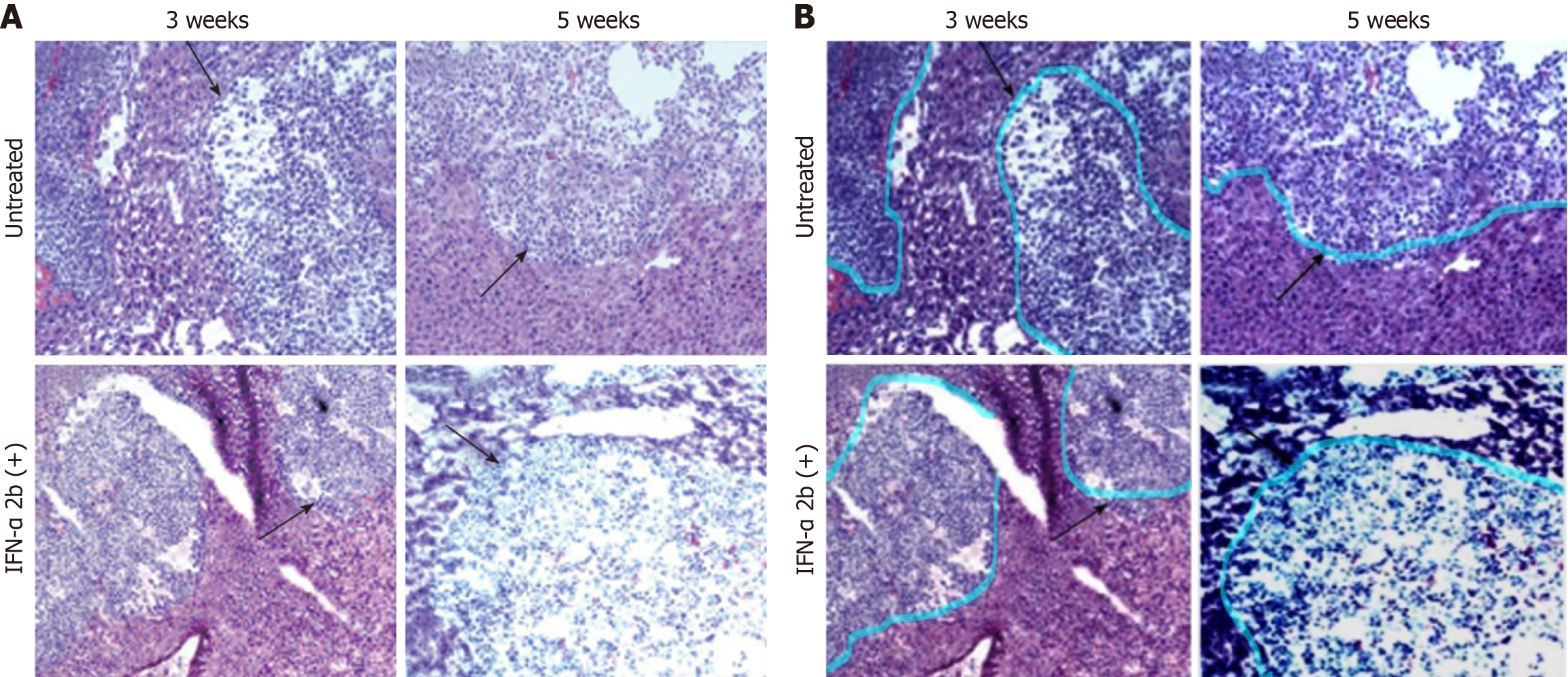
Core Tip: This correction is to: Hazari S, Hefler HJ, Chandra PK, Poat B, Gunduz F, Ooms T, Wu T, Balart LA, Dash S. Hepatocellular carcinoma xenograft supports HCV replication: a mouse model for evaluating antivirals. World J Gastroenterol 2011; 17 (3): 300-312, PMID: 21253388 DOI: 10.3748/wjg.v17.i3.300. The purpose of figure 9 in this article is to show that interferon treatment does not reduce tumor growth. The histology panel shown in weeks 2 and 4 were duplicated. As the corresponding author, I am submitting a corrected figure. This correction is requested to ensure the accuracy of the information provided in this article. This correction does not impact the conclusion of the manuscript.


CORRECTION

This is a correction to: Hazari S, Hefler HJ, Chandra PK, Poat B, Gunduz F, Ooms T, Wu T, Balart LA, Dash S. Hepatocellular carcinoma xenograft supports HCV replication: A mouse model for evaluating antivirals. World J Gastroenterol 2011; 17(3): 300-312 [PMID: 21253388 DOI: 10.3748/wjg.v17.i3.300].

This article[1] was published in 2011. It came to my attention that figure 9 panels shown weeks 2 and weeks 4 were duplicated. As the corresponding author, I am submitting a corrected figure (Figure 1). This correction is requested to ensure the accuracy of the information provided in this article. This correction does not impact the conclusion of the manuscript.

Figure 1
Figure 1 Histological evaluation of liver sections after hematoxylin and eosin staining indicates that interferon-α treatment did not cause tumor necrosis or reduce the size of tumor nodules in the SCID mice liver. A: The upper panel shows the untreated mouse liver sections after 3 and 5 weeks of tumor development. The black arrows indicate the hepatocellular carcinoma (HCC) tumor in the mouse liver. Similarly, the lower panel shows the interferon-α-treated mouse liver sections with HCC tumor at different time points (10 × magnification); B: Interferon a treatment did not reduce the size of the HCC tumor in the mouse liver. An improved version of the histology images was prepared by enhancing contrast, brightness, and color balance. The upper panel shows the untreated mouse liver sections after 3 and 5 weeks of tumor development. The pseudo-color boundary indicates the HCC tumor in the mouse liver before interferon a treatment. Similarly, the lower panel shows the interferon-α-treated mouse liver sections with HCC tumor at different time points (10 × magnification).
ACKNOWLEDGEMENTS

We thank the reviewer for these constructive comments.

Footnotes

Provenance and peer review: Unsolicited article; Externally peer reviewed.

Peer-review model: Single blind

Specialty type: Gastroenterology and hepatology

Country of origin: United States

Peer-review report’s classification

Scientific Quality: Grade C, Grade C

Novelty: Grade C, Grade C

Creativity or Innovation: Grade C, Grade C

Scientific Significance: Grade C, Grade C

P-Reviewer: Zhou SW S-Editor: Li L L-Editor: A P-Editor: Zhao S

References
1.  Hazari S, Hefler HJ, Chandra PK, Poat B, Gunduz F, Ooms T, Wu T, Balart LA, Dash S. Hepatocellular carcinoma xenograft supports HCV replication: a mouse model for evaluating antivirals. World J Gastroenterol. 2011;17:300-312.  [RCA]  [PubMed]  [DOI]  [Full Text]  [Full Text (PDF)]  [Cited by in CrossRef: 4]  [Cited by in RCA: 6]  [Article Influence: 0.4]  [Reference Citation Analysis (0)]