Copyright
©The Author(s) 2025.
World J Gastroenterol. Jun 21, 2025; 31(23): 108472
Published online Jun 21, 2025. doi: 10.3748/wjg.v31.i23.108472
Published online Jun 21, 2025. doi: 10.3748/wjg.v31.i23.108472
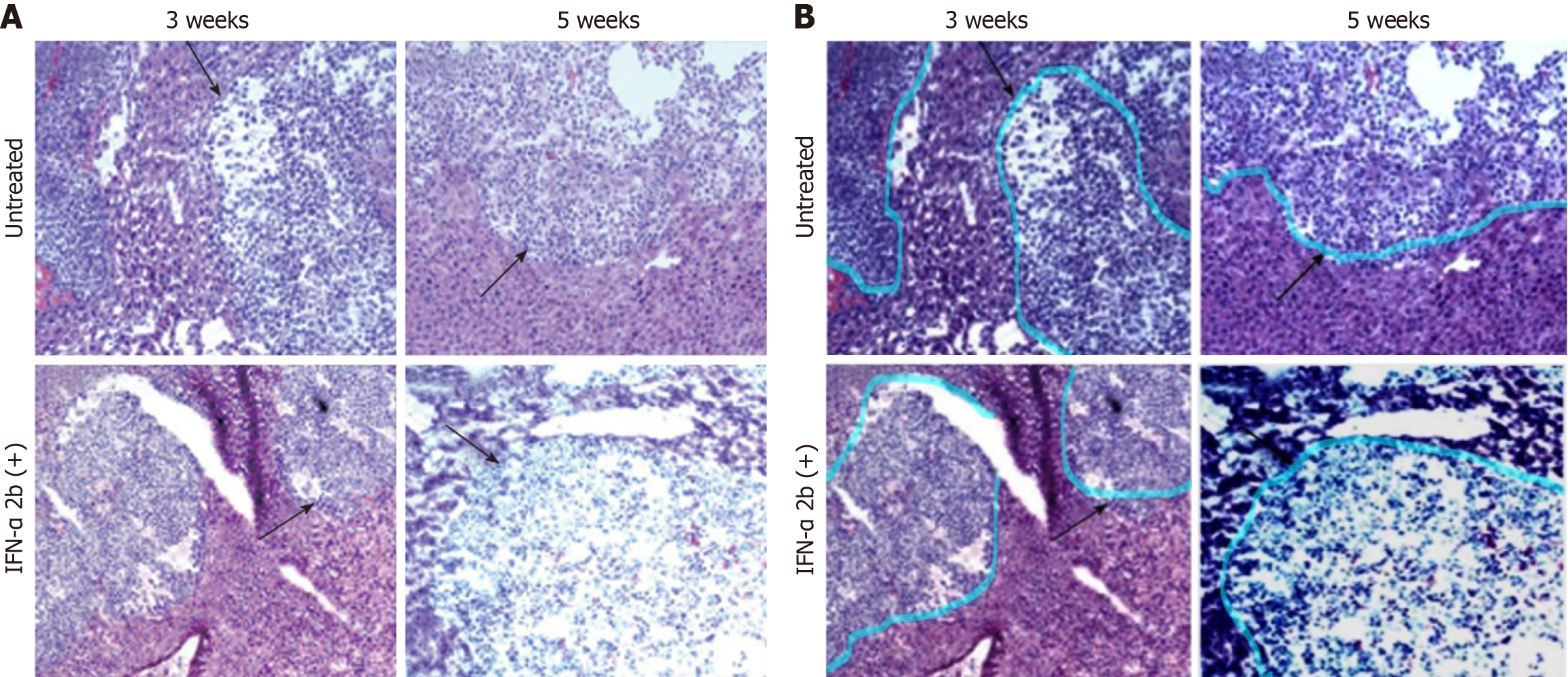
Figure 1 Histological evaluation of liver sections after hematoxylin and eosin staining indicates that interferon-α treatment did not cause tumor necrosis or reduce the size of tumor nodules in the SCID mice liver.
A: The upper panel shows the untreated mouse liver sections after 3 and 5 weeks of tumor development. The black arrows indicate the hepatocellular carcinoma (HCC) tumor in the mouse liver. Similarly, the lower panel shows the interferon-α-treated mouse liver sections with HCC tumor at different time points (10 × magnification); B: Interferon a treatment did not reduce the size of the HCC tumor in the mouse liver. An improved version of the histology images was prepared by enhancing contrast, brightness, and color balance. The upper panel shows the untreated mouse liver sections after 3 and 5 weeks of tumor development. The pseudo-color boundary indicates the HCC tumor in the mouse liver before interferon a treatment. Similarly, the lower panel shows the interferon-α-treated mouse liver sections with HCC tumor at different time points (10 × magnification).
- Citation: Dash S. Correction to: Hepatocellular carcinoma xenograft supports HCV replication: A mouse model for evaluating antivirals. World J Gastroenterol 2025; 31(23): 108472
- URL: https://www.wjgnet.com/1007-9327/full/v31/i23/108472.htm
- DOI: https://dx.doi.org/10.3748/wjg.v31.i23.108472


